

Imagine a small core biopsy from a patient being screened for a clinical trial. The tissue is limited, but the requests keep coming: H&E, several IHC markers, and perhaps NGS or another multi-omic test. Every extra section consumes tissue. Every extra day can delay trial enrollment or treatment selection.
This is where ViewsML is trying to work. The Vancouver-based company announced a $4.9 million seed round led by Wittington Ventures, with participation from Mayo Clinic, Continuum Health Ventures, and existing backers including Debiopharm. For pathologists, the more important part is not the financing number itself. It is the direction of travel: moving part of biomarker staining from a wet-lab procedure into a software layer built on the digital H&E slide.
What ViewsML says it is building
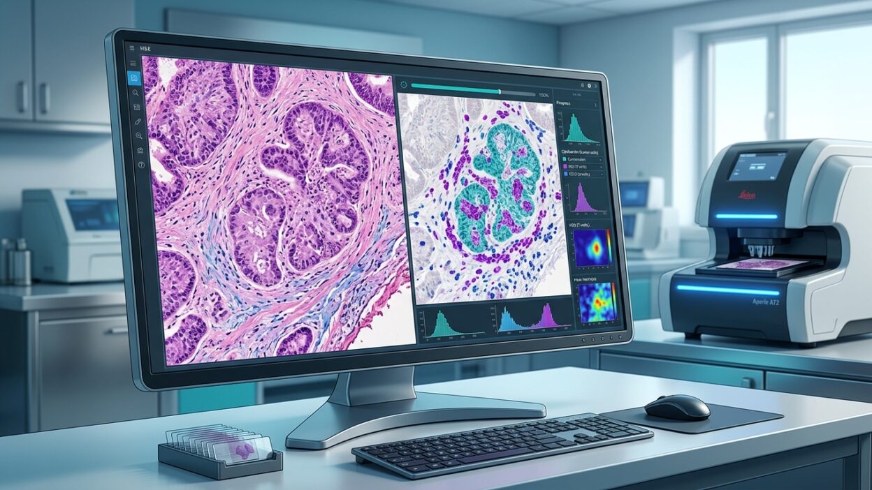
ViewsML describes its platform as a virtual biomarker library. The basic idea is that an AI model takes a digital H&E image and predicts biomarker distribution at the cellular level, producing virtual IHC or even a virtual immunofluorescence-like output. The model is not meant to stop at a case-level positive or negative biomarker call. The aim is spatial mapping: where is the protein expressed in tumor epithelium, immune cells, stroma, or vasculature?
That matters in immuno-oncology, antibody-drug conjugates, and bispecific therapies. In these settings, the question is often not only whether a marker is present. Spatial distribution and heterogeneity inside the tumor can influence patient selection, trial interpretation, and the understanding of response or resistance.
Why Mayo Clinic’s participation matters
Mayo Clinic joined the round as a new investor, but the relationship with ViewsML did not start with the financing. According to R&D World, ViewsML was already part of a broader Mayo Clinic Digital Pathology ecosystem that brings together clinical expertise, data, infrastructure, and external AI developers. The investment is separate from the clinical collaboration, but it signals that Mayo is watching virtual staining as a category serious enough for deeper testing.
For digital pathology teams, this is an important distinction. Scanning a slide is not the same thing as extracting new clinical information from it. WSI becomes more valuable when the image supports quantification, spatial mapping, triage, and links between morphology and biomarkers.
From H&E to virtual IHC
Traditional biomarker profiling usually depends on IHC followed by molecular testing such as NGS. The workflow is familiar, but it costs tissue and time. ViewsML argues that part of that process can become software: a model predicts protein localization from H&E and generates a virtual biomarker stain directly from the digital image.
If this type of tool proves itself clinically, the near-term use is unlikely to be the sudden replacement of IHC. A more realistic use is triage and stratification. The model could help teams decide which cases need confirmatory testing first, prioritize limited material, or reduce unnecessary additional sections when the biopsy is small. That is especially relevant in lung cancer, GI biopsies, and metastatic samples where the tissue barely covers diagnosis plus molecular workup.
How this differs from many current pathology AI tools
Much of pathology AI focuses on detection or classification: cancer present or absent, grade, subtype, or broad biomarker status. ViewsML is aiming for a more granular layer, with per-cell prediction and spatial context. That places the discussion closer to spatial biology than simple image classification.
The company also emphasizes virtual multiplexing. Instead of physically staining multiple biomarkers on the tissue, the system can generate several virtual layers and stack them computationally. If the approach becomes reliable, biopharma teams could study the tumor microenvironment from existing samples without repeatedly cutting tissue or running expensive staining panels.
What pathologists need before trusting virtual staining
Excitement is not enough. Any virtual staining model needs strict validation against clear ground truth. Pathologists need to know which sample types were used for training, which tumor types are covered, which scanners were involved, how fixation and staining variation affect performance, and whether the model holds up across laboratories.
The output also needs to be reviewable. Is it a probability map? Can the pathologist inspect the cells that drove the prediction? Are thresholds clear? When should confirmatory IHC still be ordered? These are practical questions, not academic details.
There is also a regulatory boundary. Use in biopharma research or clinical trial enrichment is not the same as direct use in routine diagnostic sign-out. ViewsML’s news is best read as a step toward assisted workflows, not as an immediate replacement for IHC or the pathologist’s judgment.
Why this matters for our laboratories
For laboratories planning digital pathology, the message is simple: scanning alone is not the endpoint. WSI becomes more useful when it supports tools that conserve tissue, shorten waiting time, and guide the next test more intelligently. That does not mean every lab needs virtual staining tomorrow. It does mean digital infrastructure should be built in a way that can support these tools later, with consistent image quality, good metadata, and a clear link between the image, diagnosis, and molecular results.
The ViewsML and Mayo Clinic story places virtual biomarker staining at the intersection of digital pathology and precision medicine. If these models can prove accuracy and practical value, H&E may become more than the diagnostic starting slide. It may become the starting point for spatial biomarker prediction, while preserving real tissue for the tests that still need it.
Source: R&D World