

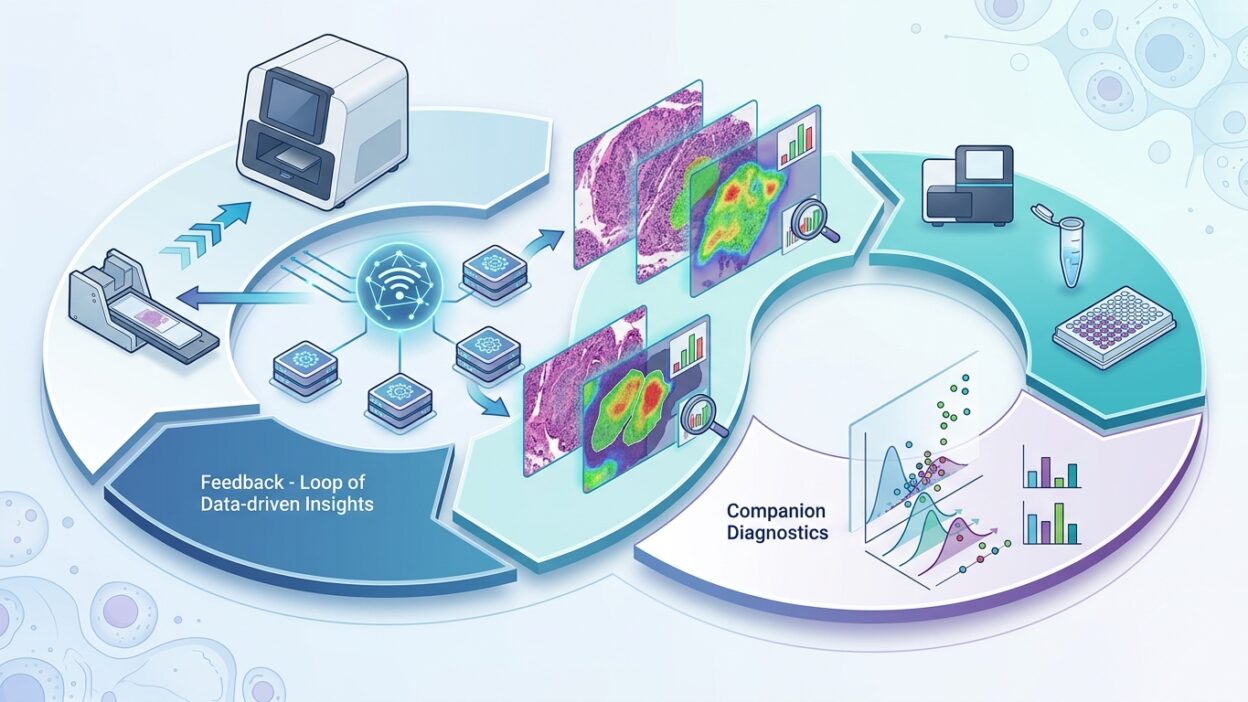
On May 27, 2026, Indica Labs announced an expansion of its companion diagnostics capabilities ahead of ASCO 2026. At first glance the news looks commercial, but it touches a sensitive point in the work of the pathologist: how tissue-analysis algorithms move from research projects or support services into tools that affect treatment selection, especially as ADC drugs expand and treatment becomes tied to marker expression and location inside the tumor.
The company connects the new offering with its HALO platforms, its experience in quantitative marker analysis, and a Precision Medicine team led by Doug Bowman. The published text says the team will cover several stages of CDx development, from discovery and validation to regulatory support. These details matter because many AI tools in pathology do not fail at the algorithm itself. They fail in the gray zone between the algorithm, the clinical platform, the quality system, and the final report read by the oncologist.
Why does this matter to pathologists?
CDx tests are no longer a simple laboratory add-on beside treatment. In a growing number of breast, lung, and colorectal tumors, treatment choice becomes linked to expression level, staining pattern, cellular distribution, or spatial relationship inside the tissue. In this setting, it is not enough for a company to say the model is accurate. The practical question inside the department is: who reviews the result? How does it appear in the WSI viewer? Where are the data stored? How are borderline cases documented? And what happens when the algorithm’s estimate differs from visual reading?
The Indica Labs announcement places AI inside a CDx pathway, not as a general triage tool. That distinction matters. A triage tool may help order cases or measure tumor area. CDx is usually tied to a regulatory file, a defined intended use, and clear limits on specimen type, stain, platform, and reporting method. The pathologist will need to read these limits before relying on any result, especially when the result affects eligibility for a targeted therapy or a clinical trial.
ADC raises the level of precision required
Indica Labs said the offering supports ADC drug analysis, as well as multiplex IHC, spatial analysis, and cell phenotyping. That makes sense. ADC drugs do not always treat the marker as only positive or negative. Sometimes the amount of expression matters, its heterogeneity, its distribution across tumor regions, the presence of a small population of highly expressing cells, or a pattern that differs between the primary tumor and metastases. These details are demanding in daily reading when the specimen is large or heterogeneous.
Digital analysis can help here, but it does not remove the pathologist’s responsibility. The algorithm can count cells, define regions, and produce a continuous score. But the meaning of that score depends on preparation quality, region selection, necrosis or crush artifact, inflammatory pattern, and tumor borders. The digital result should remain connected to informed histologic review, not become a number detached from the slide.
Quality and regulation before marketing
The statement refers to ISO, CE-IVDR, FDA premarket authorization, and notes that HALO AP carries CE-IVDR in Europe, the United Kingdom, and Switzerland, while it is Research Use Only in the United States and is not cleared there for clinical diagnostic use. That last sentence is as important as the rest of the announcement. A laboratory working in more than one market, or participating in international trials, cannot treat the same tool the same way in every country.
In practice, each department will need a clear separation between research use, trial use, and approved clinical use. It will need an SOP defining when a digital result is accepted, when it is manually reviewed, and when a case is considered unsuitable for automated analysis. In CDx, these details are not just administrative. They are part of treatment-decision safety.
The partnership with Leica Biosystems
The announcement links the new offering to Indica Labs’ partnership with Leica Biosystems. The practical value here comes from the possibility of narrowing the gap between stain, scan, viewer, analysis, and report. Many pathology departments struggle with fragmented tools: a WSI platform on one side, an LIS on another, and an algorithm running in a separate window. Every manual transfer increases error risk and makes validation harder.
If partnerships of this kind succeed in making CDx run inside one auditable pathway, adoption will become easier for quality teams and specialists. But the success measure will not be a polished conference demonstration. The real measure is how the tool performs on a busy day, with small specimens, variable stains, and cases that do not look like training examples.
What should be asked before adoption?
Before any laboratory works with a digital CDx offering, it should request specific answers. What specimen types are acceptable? What are the fixation and processing limits? How does the model handle scanner variation? Was it validated on data from more than one center? How is an unanalyzable case defined? Does the system track every human edit? And how does the output appear in the final report? These questions are familiar to anyone working in laboratory accreditation, but they become more sensitive when the decision is tied to an expensive treatment or a therapeutic trial.
There is another point. AI-based CDx may produce more detailed measurements than traditional IHC. That is useful, but it can create an illusion of precision if the clinical cutoff is not well validated. A continuous number is not an automatic clinical judgment. We need to know how the number becomes a treatment category, and who carries responsibility for cases close to the cutoff.
A short reading of the news
The Indica Labs announcement reflects a clear direction: digital-platform companies are no longer limiting themselves to slide management or research image analysis. They are moving closer to the treatment decision through CDx, quantitative analysis, and linking outputs to targeted-drug pathways. This gives pathologists stronger tools, but it also raises the level of responsibility.
The real benefit will appear when these tools enter daily work without separating the specialist from the slide, and without reducing the report to a digital output. The pathologist remains the owner of histologic judgment. AI, when regulated, validated, and documented, can become a measurement layer that helps make that judgment more consistent in cases that are hard to standardize by eye alone.