

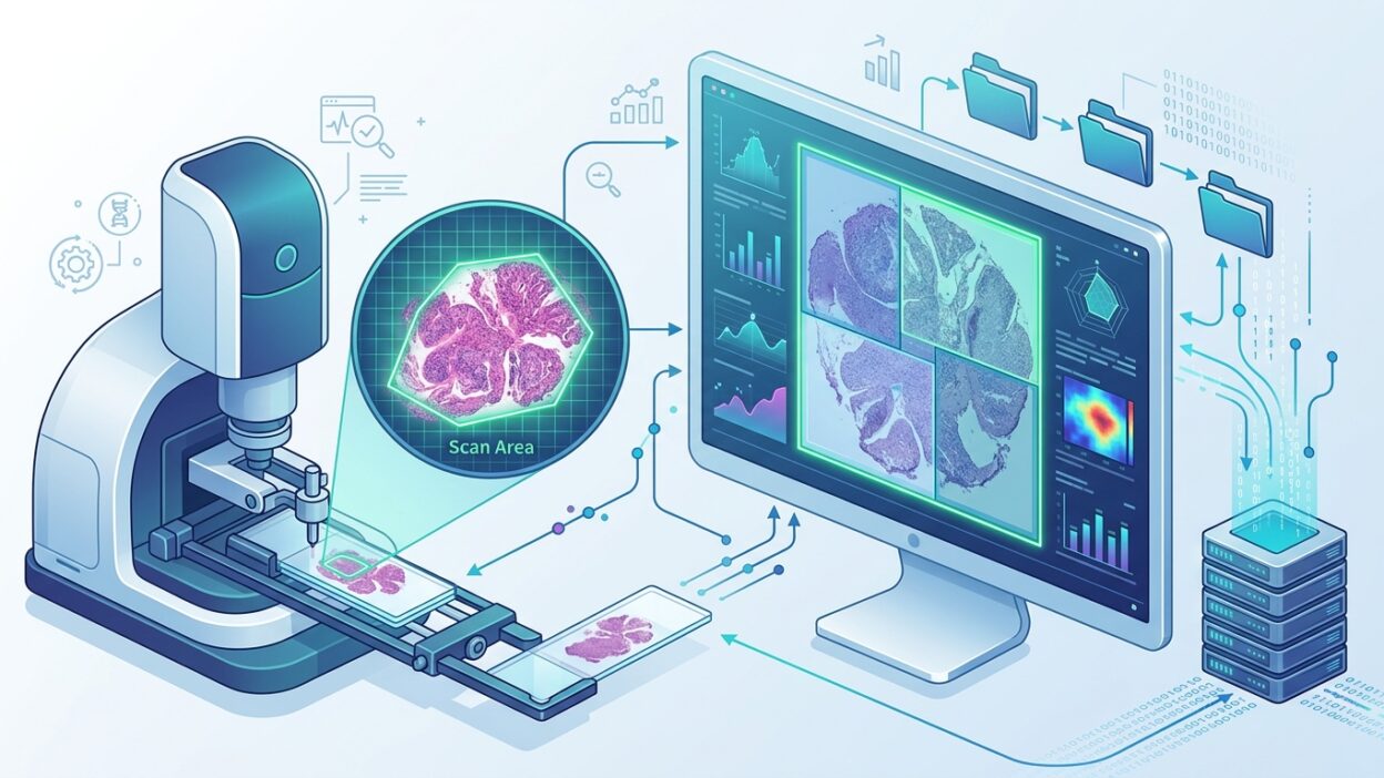
In laboratories that use whole-slide scanning, the story begins before the pathologist sees the first pixel on the screen. The scanner decides where the tissue is, builds the scan boundaries, captures the image, and then sends the file to storage and viewing systems. If the tissue detection algorithm makes a mistake at this early point, the mistake does not stay small. It becomes a scan area larger than needed, extra time on the scanner, or a heavier file in the archive.
A recent study in the Journal of Pathology Informatics, titled Comparative analysis of whole-slide scanner tissue detection algorithms, puts this issue at the center of the discussion. The article, covered by Pathology News, was written by K. Hassan Bilal with Kaitlyn Gelfant, Allyne Manzo, Victor Reuter, Meera Hameed, Matthew G. Hanna, and Orly Ardon. Its subject is very practical: how tissue detection algorithms in WSI scanners affect scan area, scan time, and file size in high-volume laboratories.
The problem starts at the tissue boundary
Many discussions about the move to digital slides go straight to image quality, viewer speed, LIS integration, or AI readiness. These are all important. But tissue detection inside the scanner itself deserves a higher place on the operational checklist because it determines how much material enters the digital production line in the first place.
The algorithm may be conservative and capture a wide empty margin around the tissue fragment. Or it may be too tight and risk leaving a small piece at the edge. In both cases, the user may not feel the problem in a single day. It becomes visible after thousands of slides: minutes accumulating on scanners, storage capacity consumed without diagnostic value, and files larger than needed slowing viewing, transfer, and backup.
This kind of detail does not look impressive in purchasing presentations. But it shapes daily work. A pathologist does not need a scanner that succeeds only with ideal slides. The laboratory needs a digital line that can handle difficult slides: small biopsies, multiple fragments on one slide, pale tissue, edge ink, bubbles, folds, and residual stain or glue. This is where the gap appears between an algorithm that looks acceptable in a demo and an algorithm that fits the routine of a busy laboratory.
Scan time is not just a technical number
Scan time is directly tied to a laboratory’s ability to finish work on the same day. When scan area increases because tissue detection is too broad, the laboratory does not only pay a storage cost. It also pays in scanner time, case waiting time, and scheduling flexibility when large batches or overnight work are involved.
In a high-volume setting, a small difference per slide grows quickly. One extra minute may not matter much across ten slides. It becomes a burden across hundreds of slides per day, especially if the laboratory has a limited number of scanners or links scanning to fixed reporting windows for remote reading. Tissue detection should therefore be part of productivity assessment, not a side note in a specification sheet.
The practical point is that the advertised speed of the scanner is not enough. The true speed appears with the laboratory’s own slides, with the tissue types, stains, and preparation habits the team works with every day. For that reason, scanners should be tested on a representative local set, not only on carefully selected vendor slides.
File size changes the economics of archiving
Broad tissue detection produces larger files. That much is obvious. Less obvious is that a larger file changes a chain of decisions: storage type, retention policy, retrieval speed, backup time, and the volume of data that may later be used for training or running analytical tools.
If a laboratory keeps WSI files for years, every unnecessary megabyte is repeated thousands or millions of times. Image compression and quality settings have a strong effect on file size, but scan area remains a major factor. Scanning empty space is not free. It adds no pathological information, but it occupies server capacity and increases movement across the network.
On the other hand, tightening the scan area without enough verification may create a worse risk: losing a small tissue fragment or an important edge of the specimen. This is not an argument for reducing file size at any cost. The goal is a controlled balance between operational sensitivity and avoiding empty space with no value. That balance is not proven by claims. It is proven by documented testing and review of failure cases.
What should the pathologist ask?
When evaluating a new scanner or reviewing the performance of an existing one, it is not enough to ask the technical team about optical resolution or system integration. The discussion needs questions closer to diagnostic work: How often does the scanner leave tissue outside the scan boundary? Which slide types confuse it? Does it handle small, scattered fragments well? Is there an automatic review step or warning when boundaries are uncertain? Can the user adjust the boundaries before or after scanning without disrupting the workflow?
These questions move the discussion from general specifications to measurable operational risks. A laboratory can build an internal validation set that includes small specimens, fatty tissue, pale IHC stains, slides with more than one fragment, and cases with irregular edges. It can then measure each scanner or setting against three points: complete tissue capture, scan time, and resulting file size.
The result does not need to be complicated. A simple table that links specimen type to rescan rate and average file size may reveal what commercial specifications do not show. More importantly, it gives pathologists a shared language with technical and administrative teams. Instead of a general objection, the problem becomes specific: this setting increases scanned area in this slide type, or this scanner needs more manual review with small specimens.
The effect on AI
Any downstream analysis tool starts from the WSI file produced by the scanner. If large areas of blank space enter the file, this may not directly harm human diagnosis, but it adds processing load and affects analysis pipelines that need to tile the image or define regions of interest. If a small tissue fragment is missed during scanning, no model can recover it later.
For that reason, tissue detection quality is not just a scanner feature. It is an early layer of quality control in the digital laboratory. It may not carry a prominent name on the project plan, but it controls the working material that reaches the pathologist, the archive, and any later computational tool.
The practical takeaway for the laboratory
The study is a useful reminder that WSI success does not depend on the final image alone. It also depends on small decisions made before the image exists: where scanning starts, where it stops, and how much non-tissue area the system allows into the file. These decisions affect the working day and the long-term storage budget.
For pathologists, the message is simple: do not leave assessment of the tissue detection algorithm to the technical team alone. Make it part of scanner acceptance, quality control, and post-implementation performance review. Digital slides do not begin when the viewer opens. They begin with the scanner’s first decision about what is worth scanning.
Source: Pathology News, with the DOI link for the article published in the Journal of Pathology Informatics: 10.1016/j.jpi.2026.100678.