

If the question around adopting digital pathology in the laboratory still comes down to diagnostic safety, this study adds a number that is hard to ignore: 99.95% concordance in clinical management when reports issued using light microscopy were compared with reports issued using digital pathology, across 2,024 histopathology cases from several specialties.
The paper was published in Histopathology under the title Digital pathology for reporting histopathology samples, including cancer screening samples. Its value is not just that it adds another concordance study. It also includes breast and bowel cancer screening samples, the area many laboratories hesitate to move into a fully digital workflow.
Study design: a practical comparison, not a theoretical test
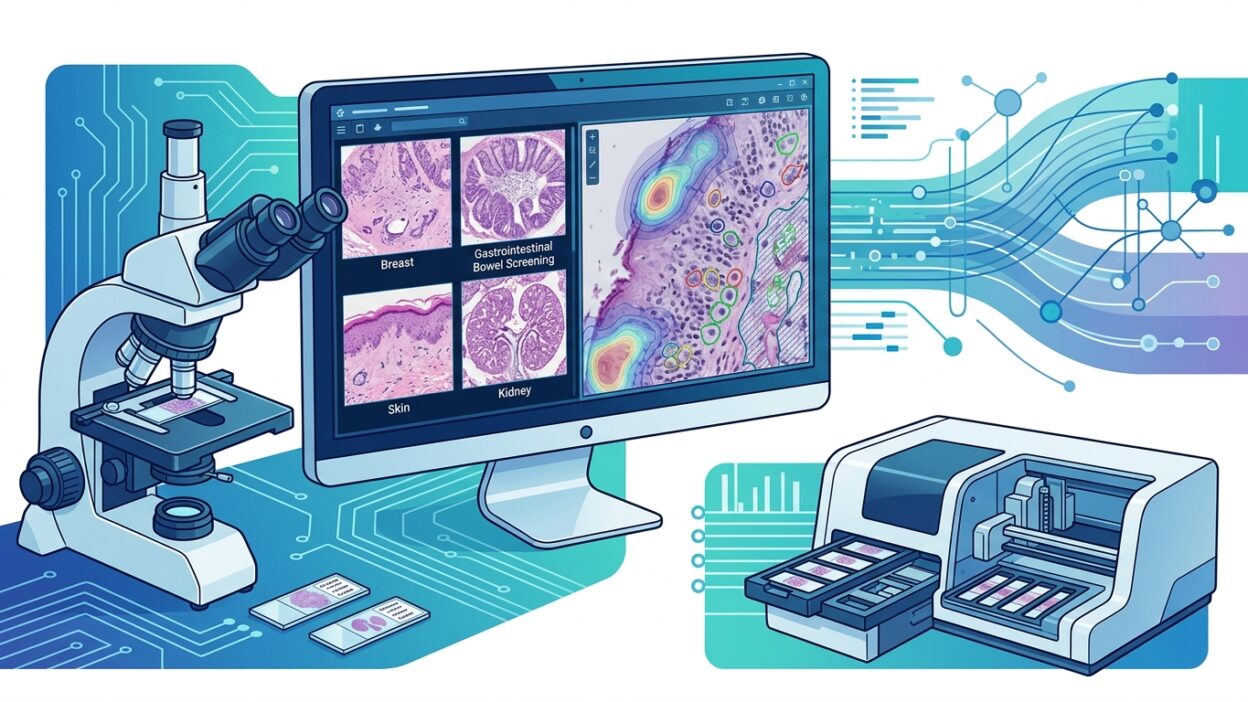
The study included 2,024 cases: 608 breast, 607 gastrointestinal, 609 skin, and 200 renal cases. Within this set, there were 207 samples from the breast cancer screening programme and 250 samples from bowel cancer screening. This matters because screening samples carry higher operational and regulatory sensitivity than routine cases, and even a small difference in classification can affect the patient pathway or programme performance.
Each case was read by light microscopy and by digital pathology, with randomized order and a six-week interval between the two readings. Sixteen pathologists took part across four specialty groups. The researchers used the concept of concordance in clinical management, meaning that the two diagnoses were either identical or that any differences between them did not change patient management.
This definition is more useful than asking only whether the two reports match word for word. In daily practice, wording differences or minor differences may not change the practical classification or treatment decision. The study therefore focused on the question that matters to the laboratory and the clinical lead: would the digital report lead to the same decision?
The main numbers
Across all cases, concordance between microscopy and digital pathology was 99.95%, with a 95% confidence interval of 99.90 to 99.97. In cancer screening samples, concordance was 98.96%, with a confidence interval of 98.42 to 99.32. The authors compared these results with a previous concordance reference of 98.3%.
When the results were broken down by specialty, the pattern was as follows: in breast cases, overall concordance was 99.40%, while in breast screening samples it fell to 96.27%. In gastrointestinal cases, overall concordance was 99.96%, and in bowel screening samples it was 99.93%. Skin and renal cases each showed concordance of about 99.99%, with high confidence intervals.
The breast screening number needs a careful reading. It does not mean that digital pathology is unsuitable for breast pathology. Rather, it suggests that this category may include areas already known to have higher interobserver variation, such as borderline lesions and classifications where histological judgment interacts with programme thresholds. The study itself notes that clinically significant differences appeared in areas known for higher observer variability, and that they occurred with both methods without a clear direction against digital reading or in favour of it.
Why this matters for managers and pathologists
Many discussions about digital transformation quickly move to scanners, storage, network speed, or viewer interface. All of those are critical, but they come after the first question: can digital reading be trusted for responsible diagnostic reporting? This paper gives a strong yes, within the limits of its design, with data that include routine samples and cancer screening samples.
The result does not mean a laboratory can switch on digital pathology by administrative decision alone. Local validation remains necessary, especially when scanner type, compression settings, staining quality, viewer software, work monitors, and specialty case mix differ. But it moves the discussion from “is digital reading safe?” to “what conditions are needed to run it safely in this laboratory?”
That is a practical distinction. A laboratory with a clear validation pathway can connect training, quality control, and exception policies to measurable outcomes. Cases that rely on fine focus, thick sections, special stains, or sensitive quantitative assessments may need stricter local rules. Still, multicenter evidence at this scale gives a stronger basis for clinical and regulatory approval.
Clinical concordance does not remove human variation
One strength of the study is that it does not present digital pathology as a way to eliminate differences between pathologists. Some differences appeared in microscopy readings and in digital readings. That reminds us that part of the problem is not the medium, but the nature of histopathological classification itself. When a diagnostic category is known to have grey zones, converting the slide to WSI will not turn it into a sharply defined category.
For that reason, this paper should be read alongside laboratory policy: when is a second review requested? When is the slide returned to the microscope? Which cases are temporarily excluded from the digital workflow? How are differences between the first reading and review documented? Successful digital adoption is not built on confidence in the image alone. It depends on a working system that knows where the image may fail and where the human observer may fail.
Practical bottom line
The study gives strong support for using digital pathology in routine histopathology reporting, extending to breast and bowel cancer screening samples. Its highest value is that it measures what matters clinically: does patient management change? In most cases, according to these data, it does not.
For laboratories still in validation, the numbers are useful as a reference when writing an internal approval protocol. For laboratories already in operation, the study is a reminder that evaluation should not stop at image quality or user satisfaction. It should reach clinical management concordance within each specialty.
Source: Azam AS et al. Histopathology. 2024;84(5):847-862. DOI: 10.1111/his.15129. PMID: 38233108.