Techcyte announced the integration of two Aira Matrix algorithms into Fusion AP, its digital anatomic pathology platform: AIRAProstate for prostate specimen analysis, and AIRAQc as a quality-control layer for whole-slide images. At first glance, the news looks like a vendor-integration announcement. For a pathology department, its value sits somewhere more practical: where AI will sit in the workflow, who will handle its alerts, and how it will affect reading time and reporting responsibility.
Embedding the tools inside an existing reading platform is different from adding a separate application at the edge of the network. Many pathology AI projects stall because the algorithm runs outside the case pathway: export, upload, wait, then return to the viewer or a PDF. Every extra step lowers real-world use, even when the algorithm is good. Bringing AIRAProstate and AIRAQc into Fusion AP puts the question in the right frame: can analysis become part of case reading rather than a parallel technical task?
Why does this matter for a pathology department?
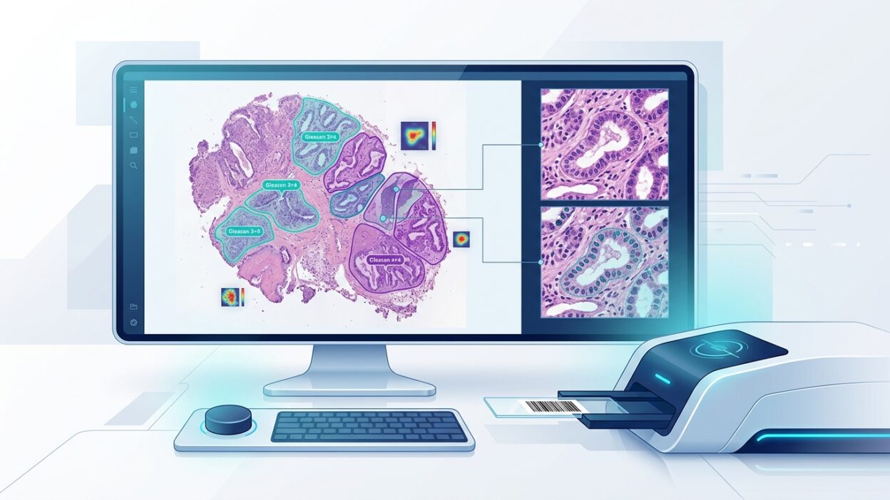
Prostate cancer is one of the more sensitive areas for any digital-analysis tool. Interobserver variation in pattern assessment, tumour distribution, and tumour length in biopsies is not a minor detail. In the clinic, these numbers affect follow-up, focal treatment, or referral into broader treatment pathways. So a useful algorithm cannot simply show a final result. It needs to help the pathologist see suspicious foci, review extent, and connect its output to the histology on the slide.
AIRAProstate, according to the published summary, is intended for automated analysis in prostate pathology. The announcement does not provide performance figures that can be used here, so it should be read as an operational integration story, not as independent clinical evidence. That distinction matters. Adopting any diagnostic tool requires local validation against the department’s specimen types, staining protocols, scanners, and working practices. Even the best vendor results do not remove the need to test the tool on local material.
The quality-control layer may be the most practical part
AIRAQc targets quality control for WSI images. This may sound less attractive than prostate analysis, but it is often closer to the daily needs of a laboratory. Focus failure, tissue folds, coverslip bubbles, unscanned areas, and staining variation can all reach the pathologist after the case has already entered the reading queue. Then the delay cycle starts: return the slide, rescan it, wait for the technical team, and reopen the case.
If the QC layer runs before the case reaches the reading list, the effect is fewer interruptions. No marketing language is needed. The pathologist wants to open a case and find a readable image. The technologist wants an early alert before defective cases accumulate. The manager wants clear measurements: how many slides were rescanned, what caused the failure, and whether the problem is linked to a specific scanner, tissue type, or shift. In that setting, a QC tool becomes more than a technical filter because it gives the laboratory operational data it can review.
Integration does not remove the need for governance
Having the algorithm inside one platform does not make the decision simpler. Successful integration raises more questions. Who triggers the analysis? Does it run automatically on every prostate biopsy or only on request? Are results shown before the pathologist reads the slide or after? How are heat maps or outputs stored in the record? Can the department trace the algorithm version that produced a result six months later?
These are not just administrative questions. In pathology, reproducibility is part of safety. If the algorithm version changes, or the scan setting changes, or the alert threshold changes, the tool’s behaviour may change. The department therefore needs a clear record linking each result to the version, scanner, date, and perhaps the original image file. Without that record, the tool may be useful in daily reading but weak during audit or case review.
There is another, more sensitive question: what should the pathologist do when the algorithm disagrees with their impression? Good use is not automatic obedience. The better role for the tool is focused review: a defined area worth another look, a small focus that may have been overlooked, or a quality alert that prevents interpretation of an incomplete image. The report remains the pathologist’s responsibility, and the algorithm should leave an understandable trace in the workflow, not a context-free number outside it.
What should be checked before purchase or activation?
The first request to the vendors should be a clear statement of the intended use. Is the tool meant for initial screening, measurement support, triage, or quality control only? General labels are not enough. The contract and workflow should define where the algorithm’s responsibility starts and where it ends.
Local validation comes next. A small but well-designed validation set is better than a broad sales demonstration. It should include easy and difficult cases, variation in sectioning and staining, samples with small tumour volume, and known quality problems. In prostate work in particular, results should be compared with readings by the department’s pathologists, followed by measurement of reading time, number of reviews, and detection of small clinically relevant foci.
For QC, the practical measurements are clearer: the proportion of slides rejected before reaching the pathologist, reasons for rejection, rescan time, and number of cases that needed repeated opening because of an image defect. These are operational indicators the laboratory can track every month. If the tool does not improve those numbers, integration into a polished platform will not be enough.
Reading the announcement as a market signal
The announcement reflects a clear direction in digital pathology: standalone algorithms lose value when they remain outside the working platform. Departments do not want a long list of scattered tools. They want a smaller number of tools that fit into reading, archiving, quality control, and result documentation without adding friction. The Techcyte and Aira Matrix partnership will matter to the extent that it reduces friction inside the department, not because of the number of logos on a product page.
For practising pathologists, this kind of news calls for a practical reading, not a celebratory one. Ask about published performance data, LIS integration, version tracking, output storage, and the local-validation plan. Above all, ask this: will the tool make the reading day clearer and faster, or will it add another screen to an already busy day?
Source: Pathology News