

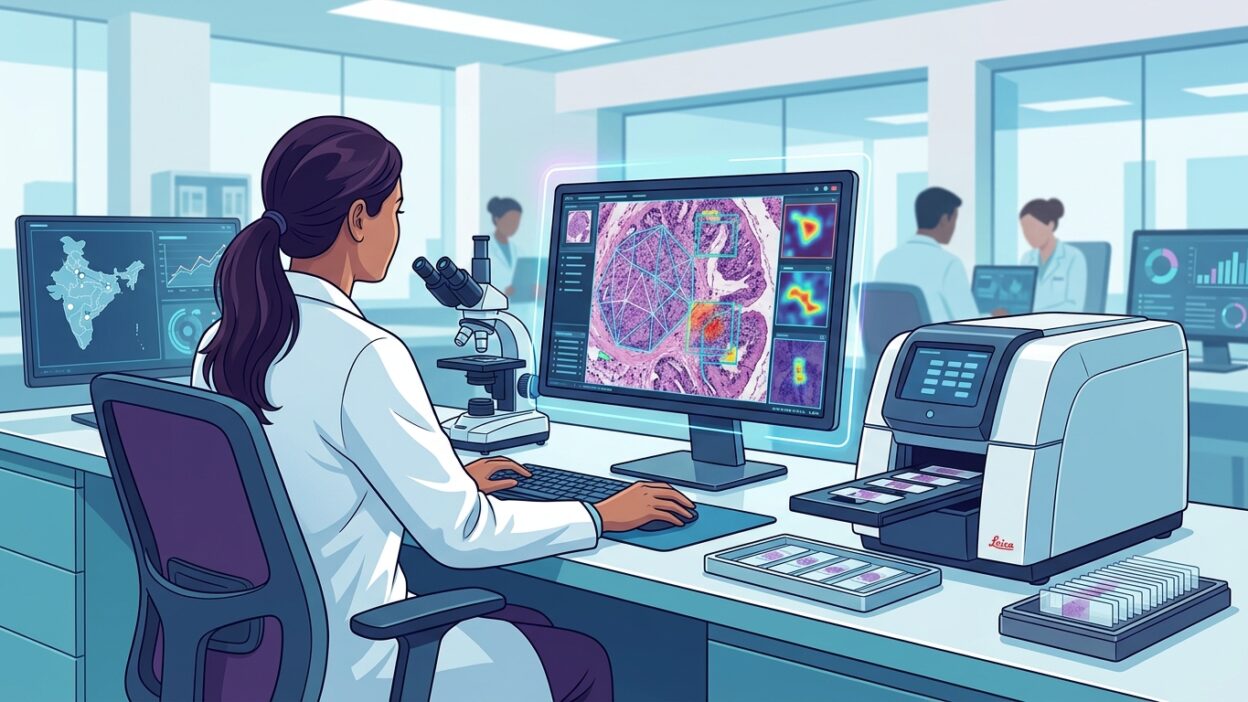
A recent review published in Journal of Pathology Informatics raises a question many pathologists outside well-funded centers know well: how can digital pathology be introduced into a large health system with uneven resources and heavy daily workloads, without turning the project into an expensive technology showcase?
The review, covered by Pathology News under the title Accelerating the implementation of digital pathology in India, was written by authors from Indian and Singaporean institutions, including Tata Memorial Centre, Rajiv Gandhi Cancer Institute and Research Center, Manipal Hospitals, Metropolis Healthcare, HCG Cancer Hospital, Roche Diagnostics India, and Roche Diagnostics Asia Pacific. That background matters. The discussion does not come only from a device manufacturer or from an isolated academic laboratory, but from several points of contact within diagnostic service.
Why the Indian experience matters to pathologists
India is not a distant special case. Population size, uneven distribution of specialists, variable infrastructure between major cities and peripheral regions, and turnaround-time pressure are familiar problems in many Arab, Asian, and African laboratories. The Indian experience therefore works as a practical mirror: if a model succeeds there under controlled conditions, its lessons can be transferred to other settings facing the same constraints.
The review argues that digital pathology adds value through WSI, remote diagnosis, and support for algorithmic analysis. This is not a wish list. The value starts when scanning becomes part of the specimen pathway, from accessioning, grossing, and staining to review, reporting, and MDT case discussion. If the digital slide remains a file separated from the LIS and IMS, workload increases instead of decreasing.
Uses closest to implementation
The review lists several applications: primary diagnosis, second opinion, telepathology, education, frozen section, algorithmic assistance, data management, and quality control. For a working laboratory, these should not be treated as one package. The most logical starting point is often second opinion and consultation cases, because the clinical return is clear and the operational risk is lower than converting all primary diagnosis at once.
Digital second opinion reduces transport of glass slides and shortens waiting time for peripheral centers. It also allows better documentation of the case pathway, particularly when several subspecialties overlap, such as breast, gastrointestinal, and lymphoid pathology. But this requires prior agreement on scan quality, file type, access permissions, and how the final opinion is entered into the laboratory’s official system.
Frozen section is more sensitive. Time is tight, section quality may be imperfect, and any network failure is felt immediately in the operating room. This use is therefore a poor starting point for a laboratory that has not yet tested scanner stability, display quality, connectivity, and the reporting pathway. It can be piloted gradually in selected scenarios, with a clear fallback route to the glass slide.
The real barrier starts before the scanner
The review lists familiar barriers: infrastructure, information technology, cost, data security, limited awareness, and concern among some staff about the effect of artificial intelligence on jobs. Reading these barriers as a missing shopping list would be a mistake. Many digital pathology problems appear before the scanner arrives: section thickness, folds, coverslip bubbles, staining variability, barcode delays, and inconsistent specimen data.
A digital slide does not hide poor preparation. Sometimes it exposes it more sharply. When the team sees the whole image on a large screen, pre-analytic problems become measurable and reviewable. That is useful, but it requires a laboratory culture that treats observations not as blame, but as a route to improving daily work.
Cost needs a more precise analysis than the scanner price alone. Storage, backup, networks, displays, maintenance, training, and LIS-IMS integration all count. Against that, there may be savings in slide transport, consultation time, archive management, and clinical meetings. A good decision is based on a local financial model, not on a generic marketing presentation.
Data security is not an administrative detail
The review calls for strong data security and the development of high-quality datasets. In practice, this means the laboratory must decide early who owns the image, where it is stored, who can access it, how access is logged, and how long it is retained. WSI images are not transient teaching images. They are part of the medical record and are tied to diagnosis, treatment, and follow-up.
When artificial intelligence is introduced, data become more sensitive. An algorithm trained on limited or non-representative samples may perform well in a paper, then degrade when staining, preparation style, or population distribution changes. India, because of its size and diversity, is a useful reminder that local performance has to be measured locally. Published numbers from another environment are not enough.
The pathologist’s role
Fear of job displacement appears in many discussions of digitization. The practical response starts by defining the physician’s role in the project. The pathologist should help choose the starting cases, acceptance criteria, validation protocols, slide display method, and final reporting standards. If the decision is left to procurement or IT alone, the result may look good from the outside and feel exhausting to use.
Training is not a single orientation session on the scanner. The team needs training in digital reading, magnification limits, handling out-of-focus areas, comparing H&E with IHC when needed, documenting snapshots, and reporting image failures. Technologists also need preparation-quality training, because a successful scan begins at the microtome, not at the server room door.
What can our laboratories learn?
The best lesson from this review is that implementation should be phased. Test one pathway, measure turnaround time, monitor image quality, calculate storage cost, and ask clinicians whether clinical decision-making actually became faster. Then expand to other services. Starting big may look good institutionally, but it raises the chance of resistance if the team feels the system added clicks and support requests more than it added value.
The review also recommends collaboration between pathologists, care providers, policymakers, and technology developers. In practice, that collaboration has to become working documents: a validation protocol, access policy, downtime plan, quality indicators, and a schedule for performance review. Without these details, digitization remains a broad label over an unstable process.
The final value of digital pathology in India, and in any similar system, will not be measured only by the number of scanned slides. It will be measured by the laboratory’s ability to provide a more precise opinion, faster sharing, a searchable archive, and data that can later support evaluation of artificial intelligence tools. The path starts with a simple question: which part of our daily work will improve if it becomes digital? When the answer is specific, the investment is easier to defend and more useful for the patient.
Source: Pathology News