

Why should pathologists pay attention to PRET now?
The Hong Kong University of Science and Technology has announced PRET, short for Pan-cancer Recognition without Example Training, as a histology image analysis system that can handle new tasks from a small number of annotated slides at inference time, without retraining or fine-tuning. The report was published by Pathology News, citing MobiHealthNews, and the technical details and results published in Nature Cancer make this closer to a practical discussion inside pathology departments than a passing technology story.
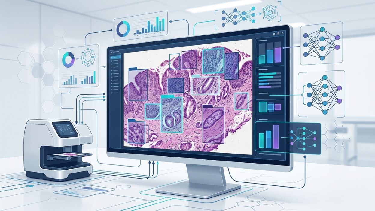
The core idea is not that the model already knows everything. What matters is that it uses local examples from the image itself, or from a few annotated slides, as visual context to perform the requested task. This kind of in-context learning is familiar in language models, but applying it to WSI raises an important question: can pathology models become less dependent on huge training sets for every tumor, every hospital, and every scanning protocol?
For pathologists, this is not a research luxury. The biggest obstacle to bringing AI tools into daily work is often the need for local data, labor-intensive annotation, long validation tests, and then model adjustment when the population, scanner, or preparation method changes. If a model such as PRET can reduce that cost, it would change how diagnostic support tools are evaluated.
What did the team do?
The university developed the system with Guangdong Provincial People’s Hospital and Harvard Medical School. According to the report, PRET was tested on 23 international benchmark datasets from China, the United States, and the Netherlands, covering 18 cancer types and several tasks, including screening, tumour subtyping, and tumour segmentation. That point matters because many pathology models look good on a narrow task, then lose value when they move to another tissue type or another diagnostic question.
The reported results are striking. The system outperformed comparison methods in 20 tasks. In colorectal cancer screening, the AUC reached 100%, and in segmentation of esophageal squamous cell carcinoma, the AUC reached 99.54%. It also recorded 98.71% for detecting lymph node metastases using only eight slides, compared with an average performance of 81% among 11 pathologists, according to the report.
These numbers are not enough on their own for clinical judgment. We still need to know how cases were selected, how grades were distributed, how many difficult cases were included, and how ground truth was defined. But the numbers give a clear reason to read the original paper, especially because the team said most validation data came from new scans in collaborating hospitals and had not been publicly available before the study. That reduces the risk of data leakage, although it does not remove the need for independent external validation.
What does in-context learning mean for slides?
According to Prof Li Xiaomeng’s explanation, the model uses local patches as contextual cues. It treats these parts as examples inside the context, then performs matrix operations that allow adaptive inference without updating the model weights. The technical phrasing matters here: we are not talking about a tool that retrains itself inside the laboratory, but about a method that lets the model benefit from a few examples during operation.
This detail brings PRET closer to real pathology needs. In a busy department, building a dedicated model for every question is not practical. But it may be possible to provide a limited number of annotated slides or documented regions for the system to use in a specific task. The difference between these two scenarios is large in time, cost, and governance.
The team also stated that PRET is not tied to a single foundation model. If that holds up in practice, it could become a layer added to more than one pathology foundation model rather than a closed product. This matters for centers with different digital infrastructure, or for centers that prefer to compare more than one model before introducing any tool into the diagnostic pathway.
What should pathologists ask before being convinced?
The first question is task boundaries. Strong performance in screening or segmentation does not mean adequate performance in borderline cases or in tumors with clear morphologic overlap. The team itself noted that distinguishing tumors with similar histologic appearance remains a current limitation. That is exactly the area where pathologists need a precise tool, not a high average number.
The second question is context quality. If PRET depends on a few examples, who chooses those examples? Is it enough for the slides to be annotated, or must they represent the full spectrum of the tumor, quality grades, staining patterns, and scanner differences? An error here may shift from the training phase to the selection of reference examples. Control of the small sample becomes critical.
The third question is integration with daily work. The report states, according to Prof Xiaomeng, that the model is open source, but it has not entered clinical operating trials and has not yet been deployed inside hospital workflows. It should therefore be treated as a strong research direction, not as a ready solution for routine diagnosis. The distinction matters, especially when companies make claims that exceed what a laboratory can verify.
Where might it be useful first?
I think the first uses will not be replacements for diagnostic judgment. They will be tasks with a clear definition and measurable outputs: triaging suspected cases, suggesting regions for review, quantification in defined tasks, or supporting segmentation when boundaries can be reviewed by a human. In these areas, the system can be tested against the current workflow without increasing clinical risk too early.
PRET may also help centers that do not have thousands of labeled slides for each tumor. Many laboratories outside major centers cannot build large enough local training sets, but they can provide a small number of high-quality reference cases. If this approach works, it could narrow the gap between data-rich laboratories and laboratories that have only recently started digital transformation.
Practical conclusion
PRET sets a useful standard for the next discussion about pathology models: not only how many slides were used in training, but whether the model can adapt to a new question from a few examples, with performance that can be checked outside the development site. That is what matters to pathologists in the end.
The report should not be read as an announcement of a tool ready for clinical adoption. A more accurate reading is that it offers a promising path to reduce the burden of data and retraining. The next step should be independent clinical validation, using difficult cases, slides from different scanners, and a clear comparison with pathologists’ performance under real working conditions.
Source: Pathology News.