

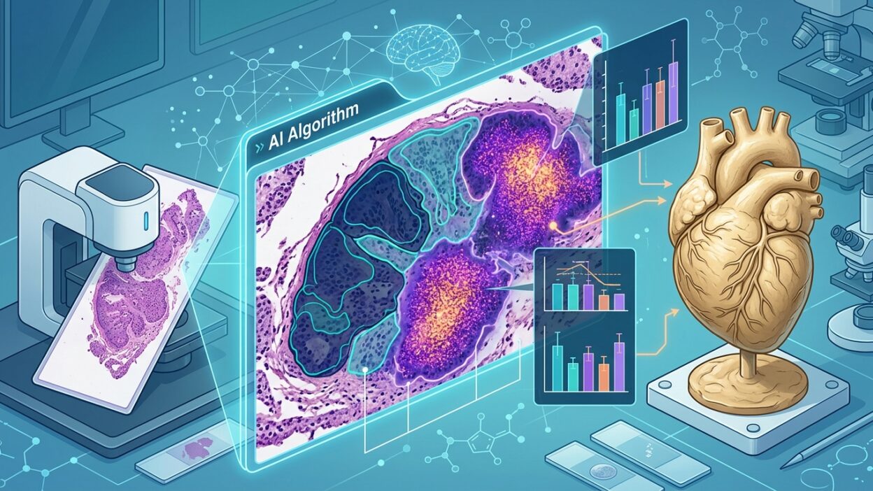
There is an idea in pathology that we often treat as final fact: histology is the gold standard. We base diagnoses on it, adjust treatment because of it, design endpoints in clinical trials around it, and then compare every new tool with it as if it were a fixed reference that does not fail.
But the article published in Beyond the Slide raises a question that deserves a pause: what if the gold standard itself was made of clay? Not because pathologists are weak, but because the instrument we use to measure disease carries its own limits.
Histology is a model, not a full copy of disease
Disease in tissue does not move in clean, ordered grades. Fibrosis, inflammation, neoplastic transformation, stromal changes, immune-cell distribution, and everything we see on a slide represent part of a continuous process. We convert that process into a Metavir stage, a Gleason pattern, or a NAS score, then treat the resulting number as if it summarizes the whole biology.
This is where the problem begins. When we compress an information-rich tissue sample into a single ordinal number, we lose details that may be the real signal. A patient within F3 may have a clear biological improvement, with less collagen or a changed inflammatory pattern, but if the visual threshold into F2 is not crossed, the trial will record that patient as a non-responder.
This is not a theoretical problem. It is an endpoint problem. A drug may be doing something important inside the tissue, while the measurement system lacks the resolution to see it.
Borderline cases expose the weakness of the system
The weakness of classification shows most clearly at the boundary between two grades. The difference between grade 1 and grade 2 lobular inflammation does not always represent a sharp biological divide. Sometimes it is a visual decision made inside a gray zone. Yet the statistical analysis in a clinical trial treats crossing that boundary as a clear event.
The same issue appears in Gleason grading. The difference between 3+4 and 4+3 does not only change the final number; it changes the meaning of risk and the patient’s pathway. A decision for active surveillance or surgery may depend on the proportion of pattern 4 seen by the pathologist. The problem is that this distinction is made in a small sample, under the influence of artefacts, processing differences, visual fatigue, and interpretive criteria that cannot be turned completely into a fixed ruler.
So it is not enough to say there is interobserver variability. The phrase has become so familiar that it has lost its edge. The more important question is where that variability sits. Often, it sits exactly at the point where a clinical or regulatory decision is made.
The biopsy itself is a small part of the truth
In liver disease, a two-centimeter biopsy may represent a tiny fraction of the organ. With diseases that have heterogeneous distribution, such as fibrosis or tumor infiltration, the place where the needle enters can change the stage. The patient may look worse or better because the sample came from a different area, not because the disease actually changed.
This matters in clinical trials. If an endpoint depends on a baseline biopsy and a later biopsy, part of the “improvement” or “deterioration” may come from sampling noise. When the treatment signal is moderate, that noise can hide the drug effect or create an effect that looks larger than it is.
MASH is a clear example of endpoint fragility
In MASH, response assessment depends on resolution of steatohepatitis or improvement in fibrosis. The NAS combines steatosis, lobular inflammation, and hepatocellular ballooning. Among these variables, ballooning is the hardest and most variable between readers, yet it feeds directly into the definition of response.
This creates an uncomfortable situation: the most subjective variable becomes part of the decision that determines whether a trial succeeds or fails. If some patients become “responders” on re-read because of measurement noise alone, the problem is in the structure of the endpoint, not only in pathologist training.
Where do digital pathology and AI fit?
The value of digital pathology here does not come from turning a glass slide into an image. The real value begins when we move from coarse visual classification to continuous quantitative measurement. Instead of stage 2, we can measure collagen area. Instead of a broad estimate of inflammation, we can calculate cell density and spatial distribution. And instead of stopping at visible tissue architecture, we can extract features associated with genomic or clinical response.
AI does not remove the pathologist’s role. It reduces noise in areas that pathologists themselves know are tiring and unstable. AIM-MASH from PathAI is a good example of this direction. Its value comes from trying to stabilize a variable part of the measurement tool, especially a variable such as hepatocellular ballooning, rather than from offering a new endpoint.
This distinction matters. Discussion of AI in pathology should not remain trapped in the question, “Does it diagnose like a pathologist?” The more mature question is: can it make measurement less noisy, more reproducible, and closer to a real outcome?
Comparison with the gold standard needs rethinking
Every new tool in digital pathology is usually compared with conventional histology. That makes sense up to a point. But if the reference itself lacks precision, the comparison becomes a trap. We may reject a quantitative tool because it does not match an unstable visual score, even though it may be closer to the biology or more strongly associated with long-term clinical outcome.
Histology will remain central to our work. What we need is to define its limits clearly and stop treating it as an absolute truth. It is a model. Every model should be judged by its noise, its limits, and its predictive ability, not only by its historical authority.
Conclusion
If we want better endpoints in clinical trials, especially in MASH and oncology, we need to redefine what we ask from the slide. The slide should not only give us a grade. It should give us continuous measurements, spatial relationships, trajectories, and multivariable models that can be linked to what actually happens to the patient.
The question is no longer: has AI reached the level of the gold standard? The more precise question is: was this gold standard solid enough to judge the future by?
Source: The Gold Standard Was Made of Clay, Beyond the Slide