

Beyond the Slide published an important essay titled “The Gold Standard Was Made of Clay.” It puts a direct name to a problem many pathologists know from daily practice, but one we rarely place at the center of regulatory and research discussions: a histologic grade does not capture the full biological truth. It is an approximation. Sometimes it is very useful. Sometimes it hides enough noise to change the outcome of a clinical trial or a treatment decision.
This issue matters to the practicing pathologist before it matters to the algorithm engineer. Any digital model, and any AI tool, will fail if its only target is to mimic a measurement system that everyone knows has limits. The practical question is no longer: can the machine match the pathologist? The stronger question is: can we build histologic measurements that are more precise than the ordinal classifications we inherited?
Histologic grades compress continuous biology
Most of the diseases we assess under the microscope do not move in discrete steps. Fibrosis accumulates gradually, inflammation changes in density and distribution, and a tumor shifts its architecture across a spectrum that does not respect artificial borders between one grade and the next. We then take that spectrum and place it into bins: Gleason, Metavir, NAS, and other systems.
These bins are necessary for clinical and research work, but they are not neutral. When a patient is close to the edge of a category, the result becomes sensitive to the section, the stain, the sampling site, and the reader’s eye. This is the edge problem in measurement: two biologically similar cases may receive different grades, while two different cases may sit inside the same category.
Pathologists know this. We see it in borderline cases, slide reviews, and consensus meetings. The essay’s useful contribution is that it connects this daily experience to clinical trial outcomes, where a grade moves from being a pathologic description to an endpoint that can determine whether a drug succeeds or fails.
When noise becomes part of the trial result
The figures cited in the essay deserve attention. In prostate cancer, expert agreement for the Gleason score can fall to a kappa of 0.3 to 0.5. That is not a small statistical detail. In the zone separating active surveillance from surgery, an estimate of pattern 4 can change the patient’s entire course.
In MASH, the problem becomes clearer. The disappearance of hepatocellular ballooning is part of the definition of response, yet it is one of the NAS components that is hardest to standardize between readers. If the least stable element defines trial success, we are not measuring the drug effect alone. We are measuring the drug plus reader noise plus sampling noise.
The essay also points to an important estimate: up to 16% of patients may be classified as “responders” when baseline slides are reread because of reader noise alone, without a real biological change. Histology reading inaccuracy may also dilute the true treatment signal by as much as 50%. These numbers are uncomfortable, but they fit what many people see in central slide review for trials.
The sample itself is part of the problem
There is another layer that even the best reader in the world cannot solve. A 2 cm liver biopsy represents roughly 1/50,000 of the organ’s volume. In a patchy disease, the patient’s shift from F2 to F3 may depend on where the needle entered as much as on disease biology.
This does not mean abandoning biopsy. Biopsy still provides information that many other tools cannot. But it does mean we should stop treating the number that comes out of it as if it were a complete image of the organ. It is a reading from a limited site, within a system of staining, sectioning, and measurement with known limits.
In clinical trials, these limits can work in both directions. A drug with a true effect may fail because noise obscured the signal. A drug with a limited effect may look better than it is because of random differences in reading or sampling. Both possibilities are bad for the patient, the researcher, and the regulator.
What should digital pathology do?
The worst use of AI here is to build a machine that reproduces the same manual grade and then celebrate the speed. That may help productivity, but it does not address the deeper defect. If an ordinal grade compresses a large amount of information, copying it more accurately does not restore that information.
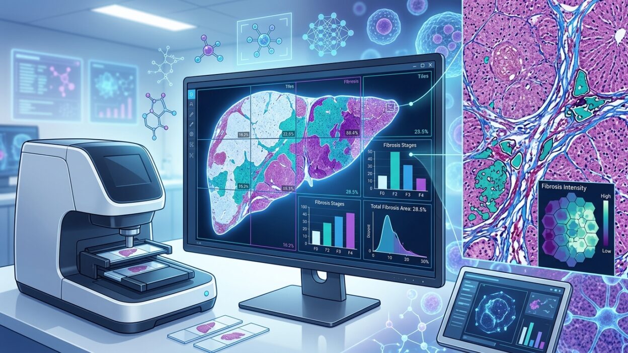
The real value begins when visual description is converted into continuous measurements: collagen area fraction, inflammatory cell density, ballooning severity, nuclear features, stromal distribution patterns, and the spatial relationships between cells, vessels, and pathologic structures. These measurements do not need to replace the pathologist’s judgment. They give the pathologist a quantitative layer that can be tested, measured again, and compared over time.
The regulatory question then becomes more exact. Instead of asking, “Did the algorithm agree with the pathologist?” we ask: does the algorithm measure a variable that links to clinical outcome better than the traditional grade? Does it reduce reader variability? Does it hold up across scanners, stains, and centers? Can it be interpreted well enough for the pathologist and the regulator to trust it?
The AIM-MASH lesson
The essay points to PathAI’s AIM-MASH as an important example. Public discussion around it has sometimes focused on a narrow question: did the FDA accept an AI-based endpoint? The more important point is that the tool was directed at a painful measurement problem, namely stabilizing the reading of highly variable elements such as ballooning in MASH.
That is the right direction. The value is that the algorithm may reduce a specific type of noise in a task that the human eye alone is poorly suited to handle when it is used as a high-stakes endpoint, while keeping the pathologist in the decision loop. The pathologist remains essential for understanding the sample, context, technical artifacts, and unexpected patterns. Quantitative measurement helps the pathologist avoid carrying alone a burden of precision that the traditional tool cannot support.
What changes in the pathologist’s work?
The practical change starts with the language we use in reports and research. When we say F3, Gleason 3+4, or a given NAS score, we should remember that the number describes a simplified model of disease. Its strength comes from communication and accumulated research use. Its weakness comes from compressing broad biology into one bin.
Pathologists therefore need to participate in designing digital metrics, not merely assess them after they are built. We know where grades fail, where readers agree, and where disagreement changes treatment decisions. That knowledge is not contained in a WSI file alone, nor in a separate spreadsheet.
The goal is not to tear down traditional histology. The goal is to put it in its proper size: a strong tool, but an incomplete one. The higher the cost of the decision built on it, the greater the need for quantitative measurement, external validation, and a direct link to patient outcomes.
Bottom line
The essay reminds us that the “gold standard” in pathology has always been a human tool built on a limited sample and a simplified classification. That does not reduce the value of histology. It makes its development a professional responsibility.
If digital pathology is to mature, it has to move beyond the race to match the reader. It has to measure what was hidden inside the grade, and test those measurements against what matters to the patient: response, recurrence, survival, and the need for a different treatment. At that point, the algorithm becomes part of a more precise science of pathologic measurement, not just another reader of the slide.
Source: Beyond the Slide