The 2D Dilemma and the Unfulfilled Promise of 3D
Throughout the history of pathology, we have relied on 2D glass slides, typically 4 microns thick. This approach effectively wastes and loses the vast majority of structural information present in the biopsy.
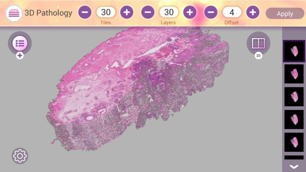
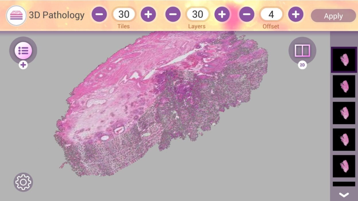
Recently, “3D Pathology” techniques have emerged, allowing us to scan the entire intact specimen without sectioning, providing a volumetric and comprehensive view of tissue architecture.
The concept is theoretically stunning, but it has encountered a frustrating practical reality: in specimens like the prostate, cancerous glands intricately intermingle with benign glands. Who has the time to precisely delineate and annotate a cancer focus, layer by layer, within a massive 3D volume?
Manual annotation here is a slow, laborious, and coarse process.
This is where a new study published in Modern Pathology steps in to solve this challenge with an ingenious algorithm named SIGHT.
What is SIGHT? (Where Magic Meets Science)
SIGHT stands for Synthetic Immunolabeling for Generative Heatmaps of Tumor.
Simply put, researchers have developed an AI system that integrates virtual staining techniques with 3D imaging.
Instead of burdening the pathologist with manual cancer detection, the algorithm digitally transforms a 3D H&E volume into a multiplexed 3D immunofluorescence image.
The system virtually stains the tissue with two Cytokeratin markers that accurately differentiate between benign and malignant glands in the prostate.
The result? A clear and interpretable 3D “Heatmap.” The cancer focus literally lights up before you amidst the healthy tissue, without consuming a single drop of chemical reagent.
The Numbers Speak: Is the Algorithm Accurate Enough?
The researchers didn’t stop at producing visually impressive images; they put the system to a rigorous test against a panel of expert Genitourinary (GU) Pathologists.
Inter-pathologist agreement among expert pathologists: Achieved an F1 Score of 0.90.
SIGHT algorithm accuracy: Achieved 0.88.
This means the system not only identifies the tumor but does so with an accuracy that nearly matches human expert consensus. The most challenging part of the process has been automated with remarkable success.
Clinical Application: Why Does This Truly Matter?
Imagine assessing the recurrence risk for a prostate cancer patient based on the biopsy’s morphological features.
The research demonstrated a pivotal point that shifted the paradigm:
When researchers attempted to predict recurrence risk using global tissue characteristics within the volume (without precise tumor isolation), the Hazard Ratio was 0.92 (i.e., no real predictive value).
However, when they used the SIGHT algorithm to precisely isolate and define “cancer regions” and performed volumetric analysis on these areas alone, the Hazard Ratio jumped to 3.57!
What does this mean clinically?
It means that the superior delineation accuracy provided by AI has made 3D data truly capable of decisively predicting who will experience disease recurrence and who will be cured. The system visualizes subtle microarchitectural changes that precede clinical relapse, which are impossible to accurately see or quantify in traditional 2D slides.
Conclusion: The Missing Piece of the Puzzle
3D tissue imaging has long been like a supercar without a steering wheel; we possess immense data, but it’s difficult to navigate and manually extract useful insights from it.
The SIGHT project represents that steering wheel. It proves that integrating virtual stains with 3D pathology is not just an academic luxury, but the tool that will make whole biopsy analysis applicable in our daily routine.
The boundaries between traditional anatomical pathology and automated analysis are blurring, and our screens are about to become smarter… and brighter!