What is a Digital Pathology Scanner and How Does It Work?
For over a century, pathologists have relied on light microscopes to examine manually stained glass slides. While this method remains the gold standard, it suffers from significant limitations: slides degrade over time, are difficult to share among physicians, and require extensive preparation time.
This is where the Digital Pathology Scanner emerged as a revolutionary tool, allowing the conversion of glass slides into high-resolution digital images that can be stored, shared, and analyzed by artificial intelligence.
—
What is a Digital Pathology Scanner?
It is a specialized device, similar to conventional scanners, but specifically designed to handle delicate tissue slides.
The device converts a stained glass slide (typically H&E or IHC) into a complete digital image called:
Whole Slide Image (WSI)
This is a virtual replica of the slide that can be magnified down to the single-cell level, just like with a microscope.
—
How does a Digital Scanner work step-by-step?
1. Slide Loading
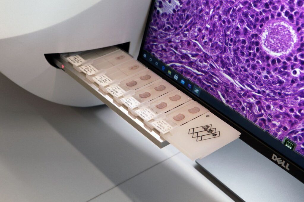
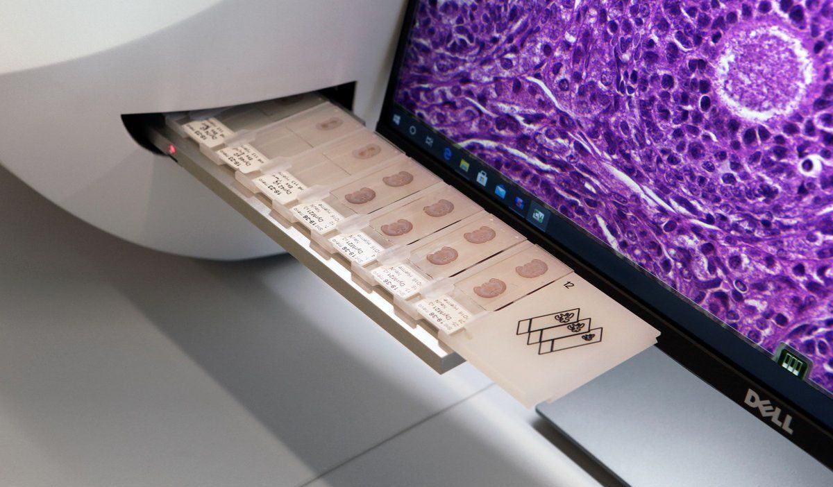
A technician places the glass slides (typically 6 – 400 slides depending on the device type) into the slide tray.
2. Microscopic Image Acquisition
The device contains microscopic lenses similar to those found in a traditional microscope.
Intense light (Brightfield or Fluorescence) is directed onto the slide.
A high-resolution camera captures small microscopic images (tiles) of different sections of the slide.
3. Image Stitching
Hundreds or thousands of these small images are stitched together using image processing software to create a comprehensive, complete image of the slide.
This image is stored in high resolution (typically in SVS or NDPI format).
4. Whole Slide Image (WSI) Production
The final result: a digital slide image that can be opened on a computer, magnified, navigated, and also analyzed by artificial intelligence algorithms.
—
Why is the Digital Scanner a revolution in Pathology?
🔹 Storage and Archiving: Instead of cabinets full of glass slides, thousands of slides can be stored on a digital server.
🔹 Remote Sharing (Telepathology): Physicians from different countries can review the same slide instantly without the need for postal delivery.
🔹 AI Analysis: Smart models can assist in cell counting, tumor size measurement, or even survival prediction.
🔹 Education and Training: Students can easily view very rare slides, instead of relying on expensive microscopes.
🔹 Quality and Speed: Reduces manual preparation errors and accelerates the diagnostic workflow.
—
Types of Digital Scanners
Brightfield Scanners: For stained slides such as H&E and IHC.
Fluorescence Scanners: For slides labeled with multiple fluorescent markers.
Hybrid Scanners: Combine Brightfield and Fluorescence capabilities.
High-Throughput Scanners: Capable of scanning hundreds of slides per day (suitable for large laboratories).
—
Conclusion
The Digital Pathology Scanner is no longer a luxury but has become an essential part of the future of pathology. It transforms the glass slide into a digital version that can be viewed anywhere, anytime, with the potential to leverage artificial intelligence to enhance diagnostic accuracy.
This technology means:
Faster diagnosis
Easier sharing
Better education
And most importantly: more accurate and efficient patient care