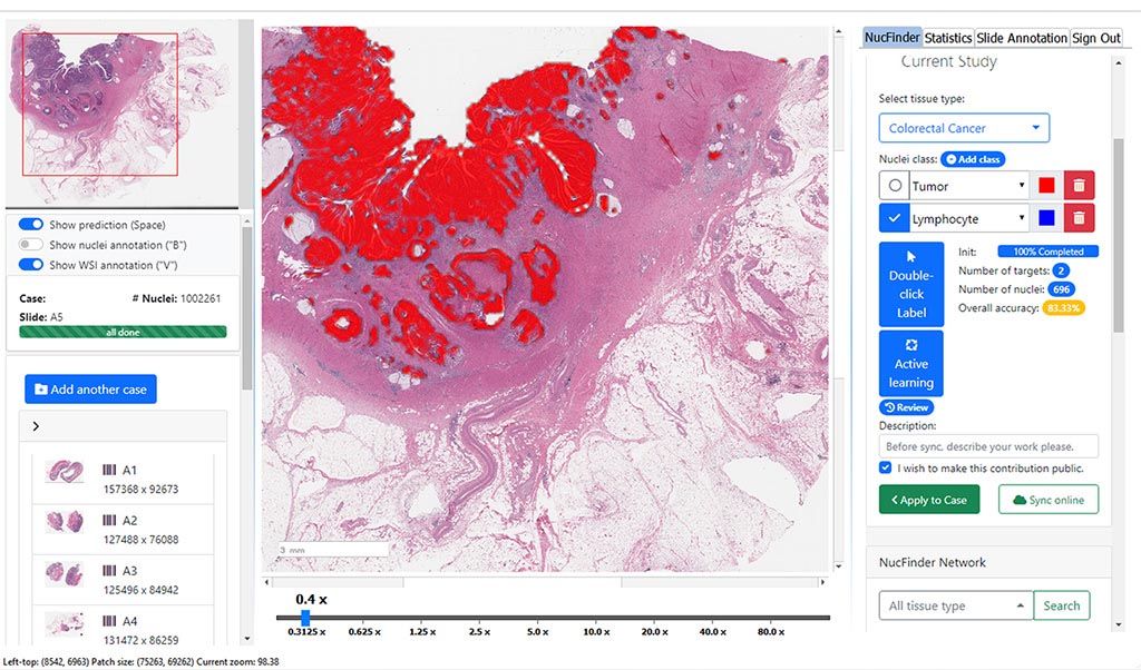
Over recent years, the term Artificial Intelligence (AI) has become extremely popular, especially with the rise of conversational systems like ChatGPT. When we talk about AI in Pathology, some colleagues might mistakenly believe it simply means:
📸 Taking a picture of a pathological slide with a mobile phone and then sending the image to an application like ChatGPT to get a diagnosis.
However, this is a completely false assumption, and in this article, I will explain why.
—
First: What is ChatGPT and what is its scope?
ChatGPT is a Language Model trained on vast amounts of text data from the internet.
Its primary function: To process words and language. It can write texts, answer questions, translate, or summarize articles.
This means it deals only with text and does not possess specialized medical expertise regarding tissue slides or pathological images.
—
Second: How does AI in Pathology differ from ChatGPT?
AI in Pathology is not a “conversational model”; rather, it consists of specialized Computer Vision models. These models are trained on:
Hundreds of thousands or millions of digital tissue slides (Whole Slide Images).
Patient data meticulously classified by expert pathologists.
Very subtle tissue patterns that cannot be discerned from a small image or a phone snapshot.
The fundamental difference:
ChatGPT = Linguistic AI (processes text).
Pathology models = Visual AI (processes digital tissue images).
—
Third: How does AI work in Pathology in practice?
1. Converting the slide to a digital format
The glass slide is placed in a specialized device called a Slide Scanner.
This device performs a complete scan of the slide with microscopic precision, producing a high-quality digital image called a Whole Slide Image (WSI).
2. Viewing the digital slide
The slide is opened on a computer, where the physician can navigate, zoom in, and zoom out as if using a real microscope.
The difference is that everything is digital: no fear of breaking or losing the slide, and it can be easily shared online.
3. Running AI algorithms
This is where the specialized models come into play.
These algorithms perform tasks such as:
Identifying suspicious areas (e.g., cancer foci).
Accurately counting cells (e.g., lymphocytes or mitoses).
Assisting in determining tumor grade or receptor status.
They do not provide a ready-made diagnosis to the physician but act as a digital assistant, highlighting key details and saving time and effort.
—
Fourth: What are the practical benefits?
Faster Diagnosis: Saving the physician’s time by focusing on critical areas.
Higher Accuracy: Detecting details that the human eye might miss due to fatigue or time constraints.
Archiving and Education: Digitally preserving slides for research, education, and future review.
Collaboration: Easily sharing slides among physicians and medical centers worldwide.
—
Fifth: Will AI replace pathologists?
This is a perpetually debated question. The truth is that current AI is designed to be:
✅ A physician’s assistant (Digital Assistant)
Not a replacement.
The final diagnostic decision will always remain with the pathologist, but the presence of these tools will make their work faster, more accurate, and safer for the patient.
—
Conclusion
AI in Pathology is not ChatGPT, nor is it merely “sending an image and getting an answer.”
It is an integrated system that begins with digitizing slides, then uses specialized algorithms trained on vast datasets to understand tissues and cells.
The goal is not to eliminate the pathologist’s role but to empower them to perform their work with higher efficiency and accuracy.
💡 Thus, the future becomes clear: Pathologist + AI = faster, more accurate, and more beneficial diagnosis for the patient.