

The tissue problem in spatial biology
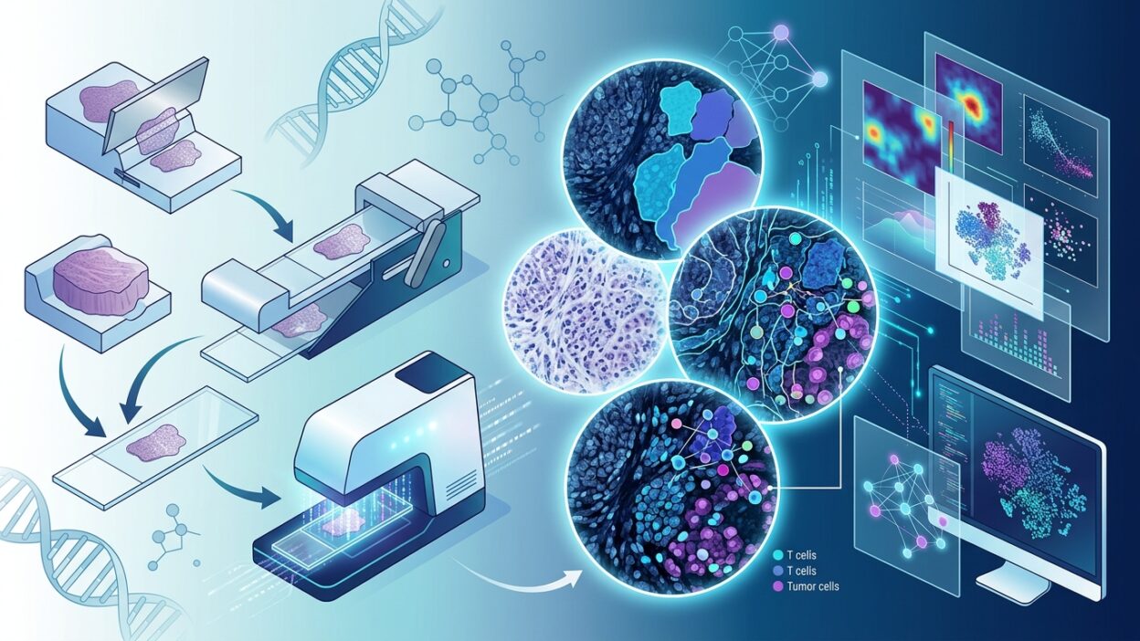
Spatial profiling is changing how we understand the tumor microenvironment, but one practical barrier remains: how can FFPE tissue sections be moved efficiently, at high throughput, onto spatial-analysis platforms? Tissue sections are fragile. Manual handling is slow and error-prone. Any tissue damage means data loss that cannot be recovered.
This is not just a technical nuisance. Every laboratory working in spatial biology knows how demanding it is to move sections from a glass slide to platform-specific assay slides, and how often tissue tears or is lost during the process. In large studies that require hundreds of sections, this becomes a real bottleneck.
What is TissuStamp?
A new paper titled TissuStamp: a novel high-throughput tissue transfer workflow for FFPE spatial biology assays, published in the Journal of Histotechnology in 2026, presents a new workflow for tissue transfer. The study comes from a team that includes Albert Velasco Abadia, Gina Benedetto, Eli Glezer, David Witters, and colleagues, the same group developing the G4X Spatial Sequencer platform.
The core idea is simple: instead of conventional manual transfer, TissuStamp provides a systematic, high-throughput method for moving FFPE sections onto spatial-analysis slides. The goal is to reduce tissue loss while increasing throughput.
Technical context
The research team works on the G4X Spatial Sequencer, a spatial-analysis platform that detects RNA transcripts, proteins, and H&E staining on the same FFPE section at cellular and subcellular resolution. The platform needs precise tissue transfer to preserve spatial registration across these different readouts.
The problem TissuStamp addresses is not secondary. In spatial multi-omics profiling, each tissue section has to be transferred to a platform-specific slide. With hundreds of samples in a single study, manual transfer becomes the bottleneck. Transfer errors mean missing data or spatially distorted data.
Why this matters for pathologists
Spatial biology is moving from academic research toward clinical use. More laboratories are considering spatial-profiling technologies for tumor characterization. Before these methods reach the clinical laboratory, their workflows need to be practical and scalable.
TissuStamp addresses one of those practical obstacles. If tissue transfer is fast, reliable, and requires less manual intervention, large spatial studies become much more feasible. That brings the field closer to a point where spatial profiling can become part of routine histopathologic diagnosis.
The next logical step is linking spatial profiling with whole-slide imaging (WSI). When RNA, protein, and morphology data are spatially registered on the same section, they can be combined with digital image analysis to provide a fuller view of the tumor microenvironment. That is where the field is heading.
Source
Velasco Abadia A, Benedetto G, Hong Z, Koh J, Duggal S, Kovács S, Vatankhah Varnosfaderani M, Kalaj M, Jeffrey-Coker B, Facchini A, Pavlich M, Senarathne WJ, Singh D, Aurelina P, Lawson MJ, Shore S, Marshall K, Ishitsuka Y, Glezer EN, Witters D. TissuStamp: a novel high-throughput tissue transfer workflow for FFPE spatial biology assays. Journal of Histotechnology. 2026. DOI: 10.1080/01478885.2026.2648709