

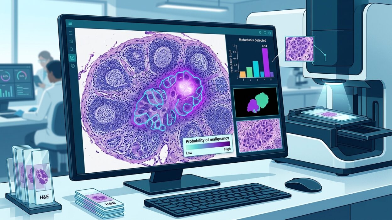
This story appeared in an earlier news cycle, but it has circulated enough to merit a second look from a professional angle. It concerns PRET, a system developed by a team led by the Hong Kong University of Science and Technology (HKUST), in collaboration with Guangdong Provincial People’s Hospital and Harvard Medical School. The main idea is clear: a model for digital slide analysis can handle more than one cancer type and diagnostic task after seeing only a very small number of labeled slides, from one to eight slides, without task-specific additional training.
For a pathologist, the value of the report is not in the marketing phrase about a plug-and-play model. The real value lies in the question PRET raises: is AI in pathology moving toward a workflow closer to expert consultation, where a few examples are enough to guide the reading, or will these results remain tied to controlled test sets that do not resemble daily workload pressure?
What did PRET do?
PRET builds on in-context learning, a familiar idea in language models, and applies it to pathology images. Instead of training a new model or fine-tuning an existing model for each cancer or task, the system receives a small number of labeled examples during inference, then uses them as an immediate reference for the target case.
The tasks described in the report include cancer detection, tumor subtyping, and tumor segmentation on slides. This range matters because many AI tools in pathology perform well in a narrow task, then lose much of their value when moved to another tumor type, a different preparation protocol, or samples from another center.
The team tested the system on 23 international benchmark datasets from China, the United States, and the Netherlands. The tests covered 18 cancer types. According to the report, PRET outperformed comparator methods in 20 tasks, and AUC exceeded 97% in 15 tasks. These are strong numbers, but they need careful reading. A high AUC does not by itself tell us where the errors occur. It does not show how the selected threshold affects sensitivity and specificity, and it does not necessarily reveal model performance on poor-quality slides, scant tissue, or mixed cases.
Lymph node metastases: the number practitioners will notice
The most prominent result in the report concerns the detection of lymph node metastases. The report states that PRET achieved an AUC of about 98.71% using only eight slides, while the average performance of 11 pathologists in the comparison was about 81%. The gap is striking, but it should not be read outside the study design.
Detection of nodal metastases is a suitable task for testing digital assistance systems because it combines a high screening burden with clear clinical risk when small foci are missed. Still, practicing pathologists know that difficulty is not evenly distributed. Some slides can be resolved quickly. Others contain tiny foci, inflammatory areas, or technical artifacts that increase the chance of confusion. The practical question therefore has to move from average comparisons to error details: where did the model fail, how large were the errors, and were its errors the kind a pathologist can easily catch within the workflow?
If the system works as a first-pass triage tool or an alert layer inside a slide viewer, its benefit may be quite different from using it as an independent reader. In the first setting, it may reduce search time in large slides and draw attention to specific regions. In the second, the requirements for validation, governance, and decision interpretation become much higher.
Few-shot models may change adoption calculations
One reason AI has entered laboratories slowly is the cost of data. Collecting tens of thousands of images, labeling them, reviewing them, and then retraining the model for each task is a long and expensive path. It does not fit every laboratory, every health system, or every rare cancer.
That is why relying on a few examples is appealing. A laboratory with a limited number of documented cases may be able to test a tool more quickly. A center in a resource-limited setting may not need to build a large dataset before it can start assessing model utility. But appeal is not enough. The few examples themselves must be representative. One poorly chosen slide may steer the system in the wrong direction, and eight slides may not capture variation in preparation, histologic pattern, and scanner differences.
This places a new responsibility on the pathologist. The role will extend beyond pressing the start button to choosing reference examples, defining acceptable cases, and setting the limits of model use. A tool that learns from a few examples makes the quality of those examples part of the quality of the final decision.
What should be required before clinical use?
Before considering the adoption of a model such as PRET, I see local testing as non-negotiable. Each laboratory needs to know how the system performs on its own slides, not only on a benchmark dataset. Differences in fixation, sectioning, staining, scanning, and image compression may change behavior enough to affect trust.
The second requirement is error analysis. Averages are not enough. We need examples of cases in which the system failed, the size of foci it missed, and the tissue types or artifacts that raised false positives. Without that, the overall number may be useful for a scientific presentation, but it is not enough to change workflow.
The third requirement is the tool’s place inside the laboratory. If it is used for slide triage, its effect on reading time, the number of regions reviewed by the physician, and the proportion of cases requiring additional return should be measured. If it is used to suggest a subtype or tumor segmentation, it should be tied to clear standards for human review and documentation in the report.
A balanced reading of the report
PRET points to an important direction in digital pathology: models that depend less on lengthy task-by-task training and make better use of a small number of reference examples. This may fit especially well with frequent, time-consuming tasks such as lymph node examination, or tasks in which large datasets are difficult to collect.
But the strength reported in the study does not remove daily questions. Does the model keep its performance on slides from small laboratories? Is it affected by scanner type? Can it handle imperfect staining? Does it give the physician an understandable decision map, or only a numeric result? These questions will determine the system’s value in the laboratory, not the headline alone.
The professional conclusion is simple: PRET pushes us to raise the quality of the questions we ask of AI tools while keeping the pathologist at the center of the decision. The published numbers are strong, and the lymph node metastasis task deserves follow-up, but the path to clinical use runs through strict local validation, transparent error analysis, and a clear definition of the physician’s role at each step.
Source: Pathology News