

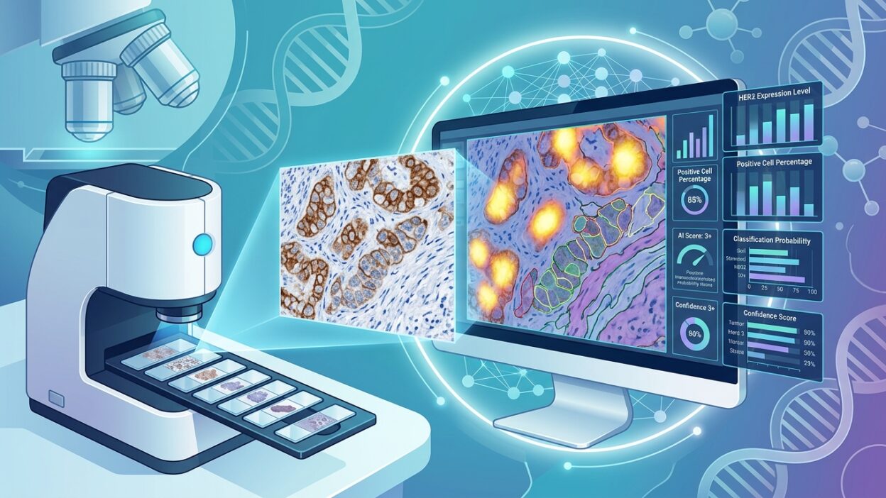
Why does HER2 in biliary tract cancer need this much scrutiny?
Today’s news pick was relatively straightforward. Among technical papers and general updates, the Pathology News report on using artificial intelligence to standardize HER2 assessment in biliary tract cancer stands out because it touches a practical diagnostic question: how do we turn an IHC stain from a variable reading into a more stable treatment decision?
The report covers a study from CHA University School of Medicine in South Korea on AI models for HER2 assessment in BTC. The context matters. HER2-targeted therapies have already entered the management of previously treated advanced biliary tract cancer, so HER2 assessment is no longer a side detail in the pathology report. The result may directly affect a patient’s eligibility for treatment.
The problem, however, is not in the obvious cases. When staining is strong and diffuse, an experienced pathologist rarely needs digital help to make the call. The challenge appears with low expression, heterogeneity, or a small sample that does not represent the tumor as well as we would like. In these cases, the distance between 1+ and 2+, or between focal positivity and clinically meaningful expression, becomes a space where readers can disagree.
The real value is not speed alone
Much of the discussion around AI in digital pathology quickly slides toward saving time. That helps, but it is not the main point here. In HER2 specifically, the stronger value is reducing inter-reader variation, especially in the zone where the pathologist is making a decision under the pressure of a limited sample, uneven staining, or tissue background that makes membrane assessment harder.
If the system can provide a consistent measure of staining intensity and cell proportions across the slide, it may move from a general assistive tool to an internal quality-control layer. It does not decide on its own. But it forces us to look at things we may pass over visually: small clusters, differences between the tumor center and edge, or a proportion of cells sitting near the diagnostic threshold.
This is what matters to pathologists more than broad performance claims. A model that produces a final score without visual explanation or a map of contributing regions will not be persuasive in a report that affects targeted therapy. A model that identifies areas of disagreement, shows the intensity distribution, and makes human review faster and more disciplined could have a place in daily practice.
Biliary tract cancer is not a copy of breast cancer
It would be a practical mistake to handle HER2 in BTC as if all previous HER2 experience transfers automatically. Intratumoral heterogeneity, sample type, inflammatory or fibrotic background, and the relative rarity of cases compared with breast cancer all make validation harder. It is not enough for the model to look good in a single internal cohort, even if the results are encouraging.
Before trusting such a tool, a pathologist needs specific details: antibody clone and staining protocol, scanner diversity, the number of low-expression cases, how thresholds were defined, and how well the model agreed with independent readers in borderline cases. Without these layers, any claim of consistency is incomplete.
Another point is just as important: what happens when the model and the pathologist disagree? In practice, this is not necessarily a failure. The disagreement may be a reason to review a specific area, request another level, or repeat the stain. But these cases need to be designed into the workflow, not left to each laboratory to improvise.
What does this mean inside the laboratory?
Introducing a model for HER2 assessment does not start with buying software. It starts with pre-analytic control. Fixation time, section thickness, staining quality, scanning settings, and slide acceptance criteria all determine whether the digital output can be trusted. The model may reveal variation, but it will not fix a poor stain or compensate for an uncontrolled workflow.
For that reason, I think the most logical first use is assisted review of unclear cases, not fully automated reading across the board. The system can triage low-expression cases, mark areas that deserve attention, and suggest a digital measurement displayed alongside the visual impression. The final decision remains with the pathologist, but the decision is supported by more consistent data.
This also matters when communicating with oncology teams. When the treating physician asks why a case is considered eligible or ineligible for targeted therapy, the answer is stronger when it rests on a documented visual read and a reviewable digital measurement. Not because the number is wiser than the eye, but because it makes the reason for the decision clearer.
Points to watch before adoption
The first thing to ask of any study in this area is external validation. A model trained and tested on slides from one environment may be affected by the staining protocol, scanner, and case distribution. This is a known risk in WSI models, and it becomes more visible when the class of interest is small or borderline.
The second point is interpretability. In HER2, a total score is not enough. The pathologist needs to see which cells the system counted, and how it handled cytoplasmic staining, background, edges, necrotic areas, and poorly preserved tissue. Without that, it is hard to defend the decision to a colleague or tumor board.
The third point is clinical linkage. Agreement between readers matters, but it is not the endpoint. The more important question is how closely the digitally supported score correlates with treatment response or meaningful clinical outcomes. A model may reduce reader variation and still add little therapeutic value if its thresholds are not linked to clinical benefit.
The practical takeaway for pathologists
This news does not mean that HER2 assessment in BTC is now solved. It means that an area we already know is variable is starting to get more disciplined measurement tools. That is a development to follow, as long as we do not confuse technical consistency with clinical judgment.
For laboratories moving toward digital pathology, HER2 in biliary tract cancer may be a suitable example for testing assistive models within a defined scope. Fewer cases, a clear treatment consequence, and a real need to reduce variation in low expression. But adoption should be built on local and external validation, not on an attractive headline or internal performance alone.
My practical view: the best place for these tools now is on the review bench, next to the pathologist, not in the pathologist’s place. If they help make borderline cases easier to discuss and document, they may be useful. If they become a final score without context, they will add a new layer of uncertainty instead of reducing it.
Source: Pathology News.