

News to read through the lens of daily practice
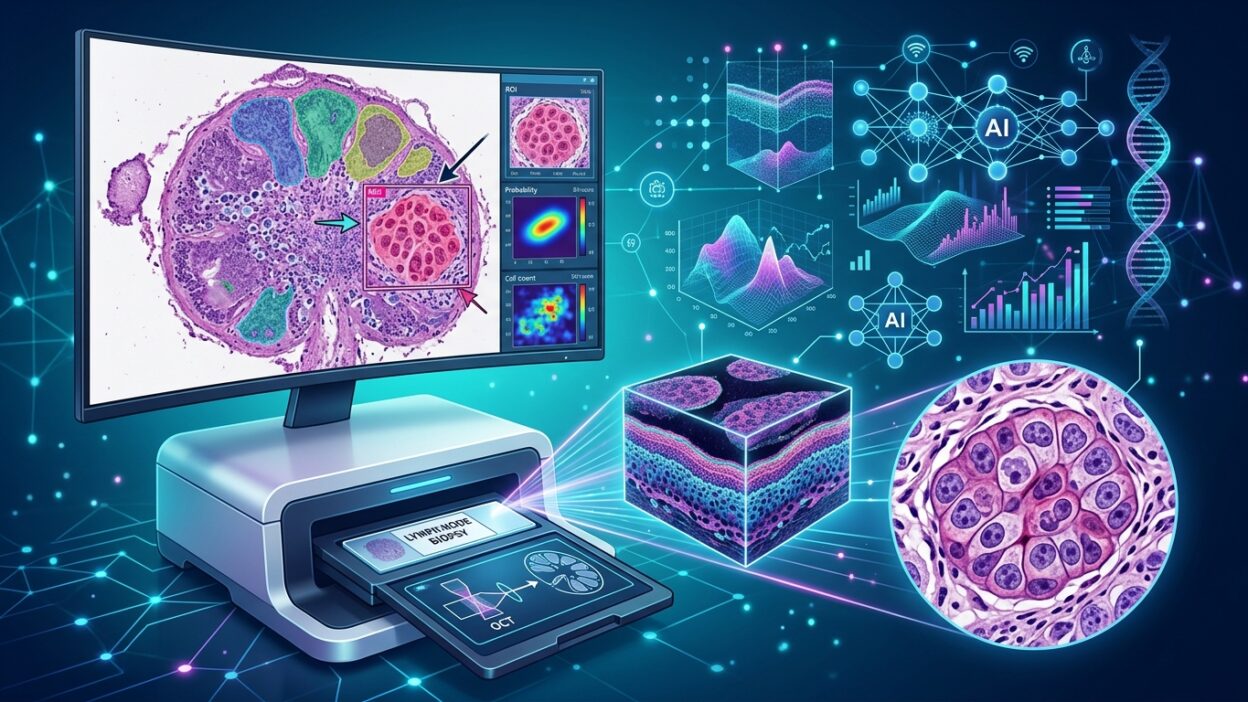
Pathology News published a summary of a recent study on using dynamic full-field optical coherence tomography, D-FFOCT, with a deep learning model to detect metastases in lymph nodes from patients with breast cancer. The title may sound technical, but the practical question is simpler: can a fresh lymph node be examined quickly enough, and with acceptable accuracy, without consuming tissue or waiting for the usual processing pathway?
The study is not describing a complete replacement for histologic diagnosis. Its value lies in a specific and sensitive point: assessing lymph nodes on fresh tissue, with an image close to histologic appearance, then converting the model output into a slide-level decision. For the pathologist, this is not only a question of speed. It is a question of where the decision sits in the surgical workflow, and who owns the decision when an algorithm enters an area that has long depended on the eye, experience, and microscopic verification.
What did the study test?
The study was conducted in a prospective two-center cohort that included 155 patients with breast cancer. The researchers obtained 747 slices from freshly bisected lymph nodes using D-FFOCT. Histologic diagnosis was used as the reference standard, and the team trained a deep learning model on 28,911 patches representing 590 slices, then tested it on 7,736 patches representing 157 slices. The results were then converted from patch level to slice level, a detail that matters more to us than the raw number at the level of a small image field.
The published figures deserve attention. D-FFOCT showed strong agreement with H&E images. Surgeons achieved a specificity of 97.10% when interpreting D-FFOCT images. The AI model achieved a sensitivity of 87.88% and a specificity of 91.94%, with an AUC of 0.899 at slice level. When a combined human-model system was used, workload fell by 75%, and specificity increased by 6.5% to reach 98.39%.
Why does this matter to pathologists?
The first point is that the study deals with fresh tissue without conventional preparation or tissue consumption. That is a practical advantage when preserving the sample matters for later tests, or when a rapid decision is needed. But it also raises a question about acceptable limits: is a sensitivity of about 88% enough in the setting of nodal metastases? In some scenarios, missing a metastasis may cost more than increasing the number of cases sent back for histologic review.
I therefore read these results as a possible triage tool, not as an independent decision. The high specificity in the combined system may help reduce work on clearly negative slices, while suspicious or discordant cases remain within the pathologist’s review. This use is more realistic than replacement talk, and closer to something that could enter a busy department without breaking the quality system.
The important detail: decision-making at slice level
Many AI studies in histologic images look strong at patch level, then lose part of their meaning when the result is converted into a clinical case. Here, the researchers linked the model output to the slice level, which makes the readout closer to daily practice. A pathologist does not sign out a report on an isolated patch. The report comes from the relationship between the specimen, where it was taken from, the clinical context, suspicious areas, and what may be outside the imaged field.
Even so, details not published in the summary remain decisive before any practical adoption: the size of small metastases, model performance in micrometastasis and isolated tumor cells, case distribution between the two centers, and the effect of different node-bisecting methods on capture. These details determine whether the tool is useful in one defined setting or transferable across different laboratories.
Where does the pathologist enter the combined system?
The finding that the combined system reduced workload by 75% is attractive, but it needs careful reading. Reducing work does not mean reducing responsibility. If a department accepts that some cases pass through an initial virtual-imaging pathway, it must define the cases that automatically return to us, the technical rejection criteria, the model’s confidence limits, and the way the decision is documented in the report or in the internal quality record.
More importantly, surgeons participated in interpreting D-FFOCT images in the study and achieved high specificity. This raises an institutional question: does the first interpretation move into the operating room, or does it remain inside the pathology department? Professionally, any tool that gives a histology-like impression and affects a treatment decision should be tied to clear diagnostic governance. Surgical participation is useful, but diagnostic accountability should not become blurred.
What do we need before clinical use?
Before this technology enters the workflow, we need independent validation on more diverse samples, clear quality-control protocols, and direct comparison with frozen section or touch imprint in the scenarios where they are actually used during surgery. We also need to know the total time from lymph-node arrival to decision, not imaging time alone. In the laboratory, the minute lost in transport, labeling, surface cleaning, and repeat imaging is part of real performance.
Another point matters just as much: the effect of the technique on the remaining tissue. The summary indicates that tissue is not consumed, which is a practical advantage, but any mechanical or organizational effect on later processing should be assessed. The method for storing images, linking them to the laboratory system, and reviewing disputed cases when model output differs from final histology also needs to be defined.
A brief professional reading
The study offers a practical direction for virtual imaging of lymph nodes: an image from fresh tissue, a model that flags suspicious areas, and a combined system that reduces work while maintaining high specificity. This direction deserves follow-up because the pain point is familiar in pathology departments, especially when time pressure meets the need to preserve tissue.
But the path to daily use runs through local validation, not through published numbers alone. If D-FFOCT enters the department, it should enter as a tool within a diagnostic pathway owned by pathologists, with clear limits on what it says and what it does not say. In that setting it may be useful: it helps the physician direct attention to the right area at the right time, while diagnostic judgment remains inside the department’s system.
Source: Pathology News, summary of a study on Virtual Histology Imaging of Lymph Nodes Via Dynamic Full-field Optical Coherence Tomography and Deep Learning to Differentiate Metastasis.