

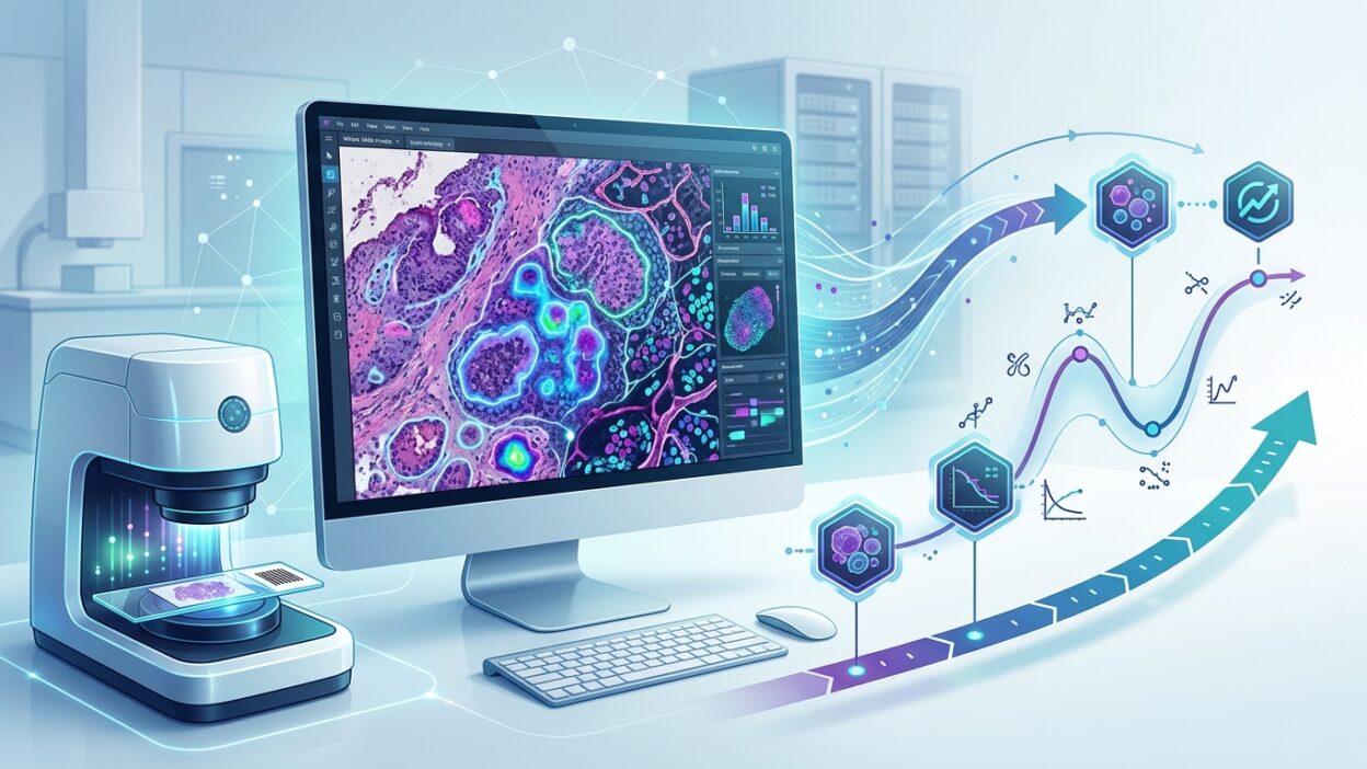
Why this discussion matters to pathologists
Beyond the Slide published an important article in a series on rethinking endpoints in the age of artificial intelligence. The central idea looks simple at first, but it reaches the core of pathology work in clinical trials: are we measuring disease as it actually occurs in tissue, or are we measuring a simplified version that fits statistical tables?
This is not just a philosophical question. In oncology practice, and in trials for liver, kidney, and immuno-oncology drugs, major decisions are turned into numbers and cutoffs. Fibrosis grade. Percentage of positive cells. Partial response. Stable disease. These terms are useful, but they do not always carry everything a trained eye sees on the slide, or everything that happens between two visits or two biopsies.
Digital pathology and artificial intelligence do not change the value of histologic judgment. They put that judgment under a stricter test: which part of our expertise should remain descriptive, and which part can be converted into a repeatable, reviewable measurement that still respects the biology?
The problem with fixed categories
For decades, medicine has classified continuous phenomena into defined categories. That was necessary. Without grades, stages, and cut points, reports cannot be standardized, patients cannot be compared, and clinical trials cannot be analyzed.
But a category is not the disease. In oncology, for example, RECIST 1.1 places a patient whose tumor burden has fallen by 29 percent in the same category as a patient whose burden has fallen by 1 percent: stable disease. A small additional difference may move the first patient into partial response. The line is statistically clear, but it is not necessarily a biological boundary.
We see the same problem in tissue every day. A fibrosis stage in the liver is not a single picture. It is spatial distribution, thickness, bridging between portal tracts, changes in lobular architecture, and an inflammatory process that may precede or accompany scarring. Reducing all of that to one number remains useful, but it loses layers of information.
This is where the article’s question comes close to daily practice: if a slide is a thin section from a three-dimensional process that changes over time, is one static reading enough to represent the course of disease? Usually not. But for a long time, it was the best tool available.
What does digital pathology add?
The practical value of digital pathology is not in turning the pathologist’s report into an automated copy. Its value appears when histologic features that are difficult to stabilize visually between readers or between centers can be measured.
In kidney disease, interstitial fibrosis and tubular atrophy can be measured as percentages, areas, and distributions rather than only as ordinal estimates. In fatty liver disease, the components of activity and fibrosis can be separated with greater consistency. In immuno-oncology, the number of immune cells in a sample is not enough. Their position relative to tumor cells, the distance between them, and their clustering pattern may carry information closer to treatment response than a simple count of positive cells.
These measurements do not replace histologic reading. They add a digital layer that pathologists must review with the same rigor they apply to a stain of doubtful quality or a nonrepresentative biopsy. A number is not true simply because it came from a model. It must be understandable, stable across scanners and preparation methods, and linked to a clinically meaningful outcome.
From amount of change to direction of travel
The strongest point in the article is the shift from asking, how much did the endpoint change, to asking, how did the course of disease move?
The difference is large. Two patients may end with the same difference between baseline and the end of follow-up, but one improved slowly and steadily, while the other improved quickly and then began to deteriorate. The final number is the same. The biology is not.
This matters in trials that use repeated samples, longitudinal digital images, or quantitative histologic endpoints. Tissue does not have to provide only a single measurement point. It can provide a direction: the speed of fibrosis progression, a change in immune infiltrate density, remodeling of stromal architecture, or a shift of the tumor toward a more resistant pattern. In that setting, the pathologist’s role becomes larger, not smaller, because the pathologist will decide whether the digital trajectory fits the disease logic we know from the microscope.
It is not enough for the model to say that risk has fallen. We need to know what changed in the tissue. Did fibrosis decrease? Did cell distribution change? Did the invasion pattern shift? Did the effect occur in representative areas, or in marginal areas that carry little diagnostic weight?
Limits of multimodal models
The article also discusses models that combine digital slides, genomics, radiology, and the clinical record. This direction makes sense because disease does not live inside one data source. A tumor is not morphology alone, a fibrotic liver is not only a scar grade, and a transplanted kidney is not a biopsy result separated from time after transplant, medications, and function.
But combining many sources raises the interpretive burden. As the number of inputs increases, it becomes harder to trace the reason for a prediction. For pathologists, this is not a side issue. If a drug trial relies on an endpoint derived from fifty variables, that endpoint must have a meaning that can be defended before a scientific committee, a regulatory body, and a patient waiting for better treatment.
The problem is not fear of technology. The problem is that an opaque endpoint may replace human subjectivity with mathematical opacity. That is not enough for publication, drug approval, or a change in clinical practice.
What should we ask of any digital endpoint?
Before accepting any AI-based endpoint in pathology, pathologists should keep a set of practical questions in view.
First: is the measurement stable across differences in preparation, scanners, and testing centers? A model that works on slides from one center may fail when section thickness, staining intensity, or scanner characteristics change.
Second: does the measurement capture a biological feature we understand? Statistical association alone is not enough. If an endpoint predicts survival, we need to know whether it depends on necrosis, fibrosis, an invasion pattern, or an unintended technical signal.
Third: does the measurement add anything to current assessment? Not every new number is useful. Some measurements add noise or repeat what the pathologist already knows. Value appears when the endpoint improves consistency, captures an early change, or separates patients whose slides look similar on routine review.
Fourth: is the endpoint linked to an outcome that matters to the patient? Digital precision should lead to a better decision, not just a better-looking figure in a research paper.
The pathologist’s role in the next phase
This discussion places pathologists at the center. Not as end users pressing a button in a digital platform, but as guardians of biological meaning. Models need someone to ask the right questions: where did you look? What did you ignore? Is the result interpretable? Is the sample representative? Did the endpoint change because of disease, or because of the laboratory?
In clinical trials, pressure will increase to use quantitative, longitudinal, and composite endpoints. Pharmaceutical companies want faster and more consistent readings. Regulators want measurements that can be audited. Patients want treatment decisions that are not lost between arbitrary boundaries. Between these groups, pathologists can prevent tissue from being reduced to a number without context.
The practical conclusion: AI endpoints should not be rejected because they are new, or accepted because they are digital. The standard should be stricter: analytical stability, clinical association, usefulness for decision-making, and a clear histologic interpretation. Only then can digital pathology move from improving workflow appearance to improving the evidence on which treatment is built.
The article works because it does not sell a technological illusion. It reminds us that the real question is not how to make measurement faster, but how to make it closer to disease as it occurs inside the patient’s body. A model cannot carry that responsibility alone.