

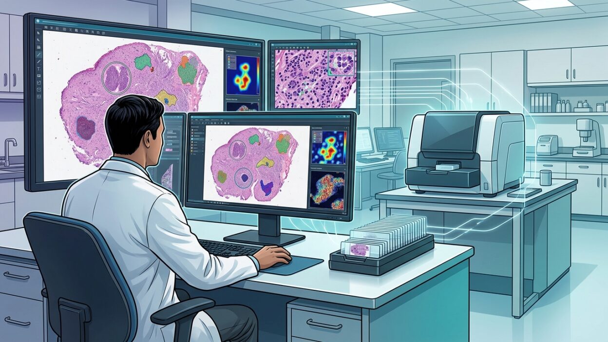
Philips’ new page on Digital Pathology is not just a marketing page for a slide scanner. Its main message is clearer than that: a laboratory moving to digital pathology is not only buying a scanner. It is changing how the slide reaches the pathologist, how the case is shared, and how algorithms enter the workflow.
For the pathologist, the important point is not the device name by itself. The practical question matters more: does it shorten the time needed to handle a case? Does consultation become easier? Can images connect with the laboratory information system, cloud infrastructure, and AI tools without creating a new burden for the team? Philips tries to answer around these points: slide scanners, an image management system, secure sharing, and connection with AI capabilities and external partners.
From slide to image, then to decision
Philips describes its aim as “accelerate the path from images to answers”. Behind that phrase is a daily problem in many laboratories: workload is rising, the number of pathologists is not rising at the same pace, and consultation between sites or teams still depends in many places on moving glass slides or sending isolated images outside the case context.
The company’s solution combines the Pathology Scanner SG60 and Pathology Scanner SG300 with the IntelliSite Image Management System. The idea is not that scanning alone solves the problem. The generated image must enter a platform where it can be searched, viewed, shared, and connected with the rest of the clinical data. This is what separates a real digital project from buying a device that sits in the corner of the laboratory.
The numbers Philips cites need a careful reading
The page says some pathologists saw efficiency improve by 15 to 20 percent per case after going digital, referring to a KLAS Research report on early digital pathology users in the United States. It also states that 35 laboratories use the Philips solution with Ibex AI, associated with productivity gains of up to 37 percent in diagnosis according to Ibex AI information and a summary published in a Modern Pathology supplement.
These numbers are useful, but they are not a ready-made promise for every laboratory. The real gain depends on specimen type, workload, scanning speed, LIS integration, network quality, staff training, and case distribution. In a laboratory where the bottleneck is slide preparation, the viewing system will not solve the problem by itself. In a laboratory with poor connectivity or no integration, the digital image may become an extra step instead of shortening time.
Secure sharing is not a secondary feature
Consultation is one of the strongest points of digital pathology. When a case is difficult or needs subspecialty input, sending a secure link to the full slide is more than technical convenience. It changes the time to decision. Instead of waiting for glass transport or compressing static images into an email, another pathologist can see the whole slide, zoom, pan, and return to different tissue areas.
Philips links this to remote work and case review between teams. This matters for hospitals with more than one site, and for laboratories that want subspecialty reporting without moving every slide to one central location. But it needs clear governance: who can open the case? How is the consultation recorded? Where is the image stored? What is the data retention policy? Technology alone is not enough if the operating policy is not written.
AI inside the workflow, not outside it
The page presents AI as part of the system, mentioning Ibex AI and integration with enterprise and cloud infrastructure. Here, a practical point matters: the value of AI in digital pathology does not appear when it runs as a separate tool that the pathologist opens only when needed. The value appears when it enters the case pathway: initial triage, marking suspicious regions, quantitative measurements, or supporting review of selected cases according to the algorithm’s scope and clearance.
At the same time, this should not be read as replacement of the pathologist. Philips itself includes important regulatory warnings: system and function availability vary by market, and some uses or versions may be for research use only in some countries. Any laboratory considering AI adoption needs to ask about local clearance, supported specimen types, algorithm limits, and how to handle cases where the pathologist’s opinion differs from the system output.
Adoption speed inside the laboratory
One point Philips mentions is that most staff in a large digital laboratory felt they became used to Philips systems within a week or less, according to a survey cited on the page. This matters because resistance to digital transition in pathology is not always rejection of the idea. Sometimes it is fear of a slow interface, loss of the familiar microscope feel, or more clicks needed to finish a case.
A successful project should start by measuring the pathologist’s daily experience, not only the number of slides a device can scan. How long does it take to open a case? Is navigation inside the WSI smooth? Is comparison between multiple levels or stains easy? Can old cases be retrieved quickly? These questions determine team acceptance more than the device specifications printed in the catalogue.
What should the laboratory ask before purchase?
If a laboratory is evaluating a platform such as Philips, it is better to request a practical demonstration on its own samples, not ideal selected cases. Small samples, large samples, H&E, IHC, slides with folds or staining variation, and cases that require comparison between more than one slide. This is where scanning strength, image quality, and ease of real work become visible.
The laboratory should also ask the company about LIS integration, file format, storage plan, access speed from outside the hospital, backup, and support for external algorithms. The Philips iSyntax file and the presence of an SDK may open a door to integration, but the laboratory needs to know what is actually available in its country and what requires additional contracts or configuration.
The takeaway for pathologists
The value of the Philips page is that it presents digital pathology as a full work platform: scanner, image management, case sharing, and AI integration. This is the right direction. The problem in many transformation projects is that they start with the device, while success starts with the workflow.
For Arab laboratories considering transition, the lesson is clear: do not ask only about image accuracy and scanning speed. Ask about case time from slide release to report sign-out, the team’s ability to consult, integration, and local support. Then digital pathology becomes a measurable operational project, not just a nice screen beside the microscope.
Source: Philips Digital Pathology page, with references cited on the Philips page including KLAS Research US Digital Pathology 2023 Performance Insights and Ibex AI information reported in Modern Pathology 2021 supplement.