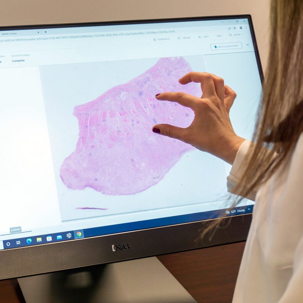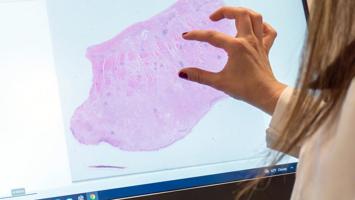
Many believe digital pathology is merely a technological luxury. Some imagine it’s reserved for giant hospitals in Europe or America, where pathologists sit before massive screens instead of traditional microscopes. However, reality tells a completely different story. If you work in a healthcare system within a developing country, you understand the extent of daily struggles in tissue diagnosis.
We need digital pathology more than affluent nations. It solves structural problems we’ve endured for decades, going beyond the idea of simply “upgrading” equipment.
A Direct Solution to Pathologist Shortages
Our biggest challenge is the severe shortage of pathologists. Entire cities or remote areas may not have a single specialist. In the traditional system, the specimen or glass slide must be transported by car to the capital or the nearest major center. Patients wait weeks for a biopsy result that could have been read in minutes.
The scanner ends this journey. A lab technician at the remote hospital scans the slide and uploads it to the system. The pathologist in the capital opens their computer, reads the slide, and writes the report immediately. This means one pathologist can cover three or four hospitals simultaneously without leaving their office.
The Nightmare of External Consultations
Every pathologist encounters complex cases and rare tumors that require a second opinion from a colleague or specialist. How do we typically manage this? We place the glass slide in a box and send it via international shipping companies to an expert abroad.
This process is slow and costly. The slide might get stuck in customs, lost, or broken during transit. Patients live in anxiety for weeks awaiting results. In the world of digital pathology, you share an image link with the consulting pathologist anywhere in the world. The expert views the specimen with full clarity and sends their opinion the same day.
Saving the Archive from Deterioration
Slide and paraffin block storage rooms in many of our hospitals lack proper standards. Heat, humidity, and dust destroy glass slides. H&E stain loses its color and fades after a few years. When a patient returns five years later with a new relapse, the pathologist requests to review the old slide only to find it unreadable.
Digital images are unaffected by time. Once a slide is scanned, patient data and their tissue history are permanently saved on hospital servers. You can retrieve them with a click of a button, with the same quality as day one.
Training the Next Generation
Resident pathologists in our countries sometimes lack exposure to rare cases. A unique glass slide can only be viewed by one pathologist at a time, and with frequent use, it may deteriorate. The digital archive transforms these slides into an open educational library. Twenty resident pathologists can study the same rare biopsy simultaneously, magnify cells, and discuss the diagnosis, without compromising the original tissue.
The True Cost
The primary obstacle raised by hospital administrators is cost. Scanners and server infrastructure involve significant expenses. However, the calculations change when we consider the full picture.
Calculate the daily cost of shipping specimens. Calculate the cost of patients traveling to seek accurate diagnoses. More importantly, calculate the cost of delayed cancer diagnoses and the subsequent expensive treatments and serious complications. Investing in digital pathology saves healthcare system funds in the long run.
Digital pathology in our countries is neither science fiction nor a luxury step. It is essential infrastructure, just like providing electricity or surgical equipment. It is the tool that shortens distances and makes accurate diagnosis accessible to the patient, regardless of their location.