

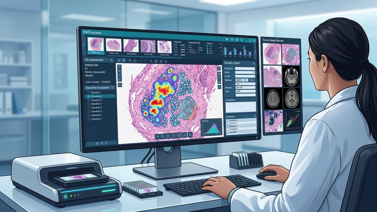
FUJIFILM Healthcare Americas and mTuitive announced on May 20, 2026, an agreement connecting the mTuitive structured reporting platform with Fujifilm’s Synapse Pathology system. At first glance, this may look like another vendor integration. In practice, it touches a daily problem inside pathology departments: what happens after the report is signed, and where does the structured data we spent time entering actually go?
The stated idea is straightforward. The structured pathology report, including CAP elements, Commission on Cancer requirements, and biomarker information, moves from mTuitive through the LIS into Synapse Pathology. After sign-out, the final report appears inside the enterprise imaging environment, not as a separate file the team has to search for later, but as part of case review alongside images and clinical data.
The problem is not only technical
Many departments now have WSI, PACS, structured reports, a cancer registry, and tumor boards. Each part works, but the connection between the parts is often weak. The pathologist signs the report in the LIS, the oncologist sees it in another workflow, radiology is in a different system, and the cancer registry still needs later extraction or manual checking.
This separation does not always look like a major failure. It appears as lost minutes, repeated entry of fields that already exist, disagreement over which version of the report each person is viewing, or difficulty collecting biomarker data that can be analyzed at institutional level. That is why connecting structured reporting to PACS matters more than the usual language of a press release suggests.
What does the connection add in practice?
According to Fujifilm’s announcement, final reports will appear automatically in Synapse after sign-out. This matters for tumor boards. When the radiology image, digital slide image, and structured pathology report sit close to one another in the working environment, case discussion depends less on emailed PDFs or screenshots saved somewhere else.
Having CAP and Commission on Cancer elements in a clear workflow also means the report is no longer only a narrative document. Structured fields such as tumor type, grade, margins, nodal status, and biomarkers can move more cleanly into clinical analysis, registries, and research. This does not remove the role of the LIS. The announcement states that the LIS remains the system of record, which is an important condition for any pathology department that wants to avoid multiple competing versions of the same report.
Biomarkers need a better pathway
In modern oncology reports, the value of the report is no longer limited to histologic diagnosis. p53, Ki-67, HER2, PD-L1, MMR, and other results feed directly into treatment decisions, and sometimes into trial eligibility or targeted therapy selection. When these data remain inside free text or an isolated system, retrieving them later becomes tiring and unreliable.
Structured reporting solves part of the problem, but it is not enough if the fields stay trapped on one screen. Linking them to the enterprise imaging environment places these fields closer to images and clinical discussion. For pathologists, this makes the quality of structured data entry visible to the whole team. A small error in one field can move quickly into a tumor board or internal analysis. The same accuracy becomes more valuable.
What about artificial intelligence?
The announcement says structured data can support clinical analytics, research, and AI applications. That line should be read carefully. The news does not claim a new diagnostic algorithm or a specific clinical performance result. The value here comes before the algorithm: organizing the data that algorithms need in the first place.
Any model that depends on WSI alone will soon face the question of how to connect the image to clinical outcome, tumor stage, biomarker status, and line of treatment. If the structured report is available in a readable and analyzable workflow, the cost of building internal datasets for review and research falls. That does not make the data immediately ready for research use. Governance, approvals, de-identification, and quality control still matter. But it reduces one of the hardest layers of the work: where is the information, and how is it connected to the image?
What should pathology departments ask before adopting this type of integration?
The first question is data responsibility. If the LIS remains the official record, the department must define what appears in Synapse, when it updates, and how post-sign-out changes are handled. Pathology reports are not always static documents. Addenda, corrections, later IHC or molecular results, and outside reviews all happen. A good integration must handle these cases without creating clinical confusion.
The second question is the design of the structured fields. Integration does not fix a poorly designed report. If templates are too long or do not reflect how the department works, physicians will resist them or fill them with the minimum required information. The real value begins when templates are precise, short where they should be, and aligned with what treatment teams and cancer registries actually need.
The third question is measurement. Before implementation, the department needs a baseline: sign-out time, tumor board preparation time, how often data are re-entered, and the rate of missing fields in structured reports. After integration, improvement or failure can be measured clearly. Without these measurements, the new interface remains a good first impression rather than proven progress.
The practical bottom line for pathologists
The Fujifilm and mTuitive agreement is not news about a new slide scanner or a new diagnostic model. Its strength is quieter: connecting the structured report to the imaging and clinical decision workflow. If implemented well, it can reduce repeated work, improve tumor board preparation, and make cancer and biomarker data easier to review and analyze.
Success will not come from the platform name alone. It will come from template quality, a clear role for the LIS, controlled post-sign-out updates, and team training so structured data become part of daily work rather than another administrative burden. For pathology departments already using WSI and PACS, this type of integration is a practical test of digital maturity: are the images merely digital, or has the reporting workflow itself become clinically usable?
Source: Fujifilm Healthcare Americas