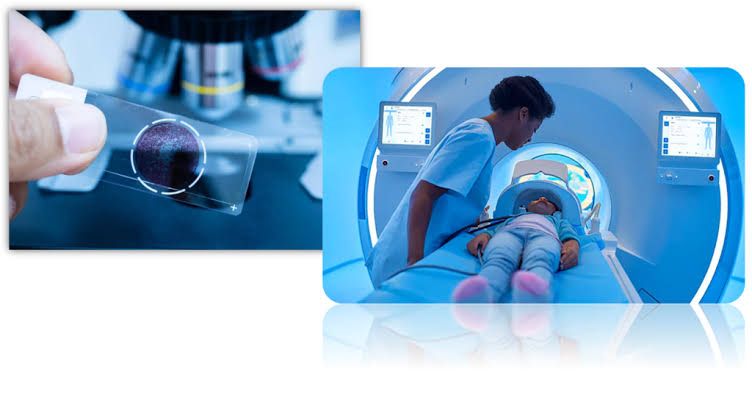
Artificial Intelligence (AI) is no longer just a futuristic technology discussed at conferences; it has become a fundamental infrastructure for modern healthcare, specifically in the field of medical imaging, encompassing both Radiology and Pathology.
The challenge we face today is not a lack of information, but a deluge of data. Hospitals daily generate terabytes of radiology images and digital slides, and traditional diagnostics struggle to process this immense volume with the required speed and accuracy.
This is where technology makes its mark: AI does not aim to replace the physician, but to serve as an “intelligent assistant” that perceives what the fatigued human eye might miss, transforming silent data into critical clinical decisions.
Why has AI become an “imperative necessity” rather than a luxury?
Medical imaging is at the heart of diagnosis. With the increasing complexity of diseases, relying solely on the naked eye has become a significant challenge. AI intervenes here to standardize criteria and automate the detection of complex patterns in both specialties:
1. In the World of Radiology: Accuracy Beyond Human Vision
Today’s AI models are trained on hundreds of thousands of X-rays, MRIs, and CT scans.
Detection of Subtle Details: Thanks to Convolutional Neural Networks (CNNs), the system can capture microscopic details, such as hairline fractures or very small pulmonary nodules that might otherwise go unnoticed.
Workflow Prioritization: The system not only reads images but “prioritizes” them. It places critical cases (such as cerebral hemorrhage) at the top of the physician’s worklist for immediate review, saving lives in emergency departments.
2. In the World of Pathology: From Subjective Estimation to Digital Precision
As laboratories transition to Digital Pathology, scanners produce gigabyte-sized slides. Manually reviewing every pixel is an arduous process.
Quantitative Analysis: Instead of approximate estimation, algorithms count mitotic cells, precisely determine tumor proportion, and classify malignant tissues.
Precision Medicine: Integrating slide images with patient history and genetic data allows AI to link “cell morphology” with “treatment response,” which is the essence of Precision Medicine.
Beyond Interpretation: How Do Algorithms Improve Image Quality Itself?
The true paradigm shift we are experiencing in 2026 is not just in “reading” images, but in “generating” them. AI-Powered Scans have revolutionized how images are captured and processed:
A. AI Reconstruction
Previously, obtaining a clear MRI image required a long time, and a crisp CT image demanded a high radiation dose.
Now, algorithms are used to “generate” ultra-clear images from less raw data (low-dose raw data).
Benefit: Significantly reduced patient radiation exposure and examination time, while maintaining diagnostic quality comparable to traditional scans.
B. 3D Visualization and Surgical Planning
We are no longer confined to 2D images. Intelligent systems can now perform “Segmentation” (organ separation) and build 3D models of tumors and organs in seconds.
This provides surgeons and oncologists with an interactive “roadmap” before entering the operating room, increasing the success rates of complex surgeries.
Standardization: Equity in Diagnosis
One of AI’s most significant features is its “Scalability.”
In traditional methods, diagnostic quality depends on the physician’s expertise and equipment quality, creating disparities between major hospitals and rural centers.
AI imposes a “unified standard.” A cancer detection algorithm operates with the same accuracy whether in a sophisticated hospital in the capital or a remote clinic. This standardization reduces human error and ensures all patients receive the same level of diagnostic precision.
A Look to the Future: From Diagnosis to Prediction
We are now in a rapidly accelerating transitional phase. AI in medical imaging is evolving from being a “diagnostic” tool (telling you what is in the image) to a “predictive” tool (telling you what will happen to the patient).
The future lies in Collaboration. Human expertise will not be replaced, but immensely augmented. Physicians who adopt these tools will transform from “image interpreters” into “clinical decision engineers,” relying on precise analyses that integrate radiology, pathology, and clinical data to improve patient outcomes unprecedentedly.
Conclusion: We are not witnessing the end of the physician’s role, but rather the dawn of a new era of “Augmented Intelligence” where technology and medicine speak a common language.