

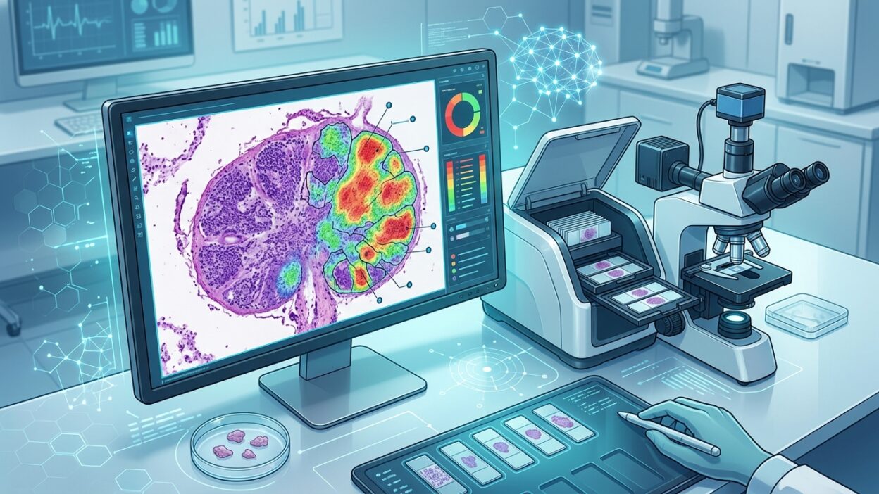
Pathology News reported an open study in Scientific Reports describing an AI model for detecting breast cancer metastases in digital lymph node slides, localizing tumor deposits, and estimating patient-level pN stage. The scientific title of the study is: Detection, localization, and staging of breast cancer lymph node metastasis in digital pathology whole slide images using selective neighborhood attention-based deep learning.
The importance of the paper does not come from a single performance number. The part that feels closer to how pathologists work is that the model does not only classify a small image patch. It combines nuclear-level reading with the surrounding tissue context. That matters in lymph nodes, where scattered tumor cells, sinus edges, inflammatory changes, and small tissue fragments can push an algorithm toward a fast decision if every patch is treated in isolation.
What did the study test?
Researchers from York University and Sunnybrook Health Sciences Centre developed a two-stream analysis system. The first stream extracts nuclear-level information through cell segmentation and classification. The second stream extracts higher-level tissue representations from the digital image. The system then uses selective neighborhood attention, choosing the neighboring patches most relevant to the patch being assessed.
This makes the decision resemble a familiar diagnostic question: are the suspicious cells convincing in their context? In daily practice, a pathologist does not look only at one cell, and does not rely only on a low-power view. The decision is built by moving between magnifications, distribution pattern, nuclear appearance, the relationship of cells to sinuses and trabeculae, and then the size of the focus and its effect on staging.
The study tested the model on CAMELYON16 and then evaluated out-of-training performance on CAMELYON17. This detail matters when reading any AI work in digital pathology. Performance inside one dataset is not enough to judge use across different hospitals, scanners, preparation protocols, and staining workflows.
The numbers that matter to pathologists
At patch level in CAMELYON16, the model reached 96.2% ± 1.5% sensitivity, 95.3% ± 2.4% positive predictive value, and an F1 score of 95.7% ± 3.1%. These are strong numbers for a task heavily affected by the ratio of small tumor foci to benign tissue, but they remain numbers from a specific testing environment and still need local validation inside the laboratory.
For tumor boundary detection, the Dice score reached 90.5% ± 2.0%, and the Jaccard index reached 82.6% ± 0.8%. This is closer to daily diagnostic need than binary classification alone. Detecting a metastasis matters, but defining its extent affects the distinction between isolated tumor cells, micrometastasis, and macrometastasis, and affects how tumor burden inside the node is documented.
At slide level, the AUC was 0.96 ± 0.01 in CAMELYON16. When the model was moved to CAMELYON17, the patch-level F1 score dropped to 87.0% ± 1.8%, and slide-level AUC reached 0.88 ± 0.03. That drop is expected and useful. It places performance closer to real-world use: models that look excellent inside one dataset may change when the data source changes.
For automated pN classification, the model recorded a kappa of 0.94 ± 0.02. If this level of performance holds in independent and local testing, the practical benefit will not be replacing pathologist review. It will be reducing the chance of missing small foci, providing an initial map of suspicious areas, and speeding review of slides with heavy tumor burden or long negative slides.
Why combining nuclei and context matters
Many WSI models treat the slide as a large bag of patches. This can work in some tasks, but it can weaken when foci are small or when there is no detailed patch-level annotation. In nodal metastasis, the difficulty is not just finding a different color. Lymphocytes, endothelial cells, artifacts, crushed areas, and weakly stained regions can look similar enough to confuse a model.
Nuclear information gives the model an input closer to histologic cytology: nuclear size, density, clustering pattern, and some differences that may not be clear in a general patch representation. Tissue context prevents the decision from becoming a search for isolated dots. When both are combined, the output becomes closer to reviewable decision support, especially if the heatmap displays the selected foci clearly.
The study also performed ablation analyses and showed that removing nuclear features or removing neighborhood attention reduced performance. That matters because it links the improvement to a defined part of the design, instead of leaving the reader with a black box that is simply described as performing well.
A practical reading before clinical adoption
For the laboratory, the practical question is where this type of model enters the workflow. The closest position is initial triage of breast cancer lymph node slides, with suspicious regions displayed and approximate extent measured. It could also be used as a second-check layer in negative cases, or as a tool for prioritizing the worklist when there are multiple nodes and multiple slides for one patient.
Clinical adoption needs more than published performance. The model must be tested on the laboratory’s own slides, including scanner differences, section thickness, local H&E, folds, bubbles, and post-treatment cases. Its behavior with ITCs, foci near the subcapsular sinus, and cases where IHC is needed to resolve uncertainty must also be known.
Another point matters just as much: how the result appears inside the image management system. If the output is only positive or negative, its value is limited. If the algorithm shows a zoomable map, reviewable borders, and a clear measurement of the largest focus, it becomes closer to an internal work tool that can be audited and documented.
Professional bottom line
This study adds evidence that digital pathology models become more useful when they approach the actual reading process: cell, tissue, context, then stage. The published numbers are encouraging, especially slide-level AUC and kappa for pN estimation, but the drop on CAMELYON17 puts cross-institution validation at the center of any adoption decision.
For daily practice, the nearest value is support for reading breast cancer lymph node slides, not independent diagnosis. If these tools enter laboratories, their success will depend on three things: a clear map for the pathologist to review, strict local validation before use, and good integration with WSI and LIS systems without adding workload.
Source: Pathology News, and the original study in Scientific Reports.