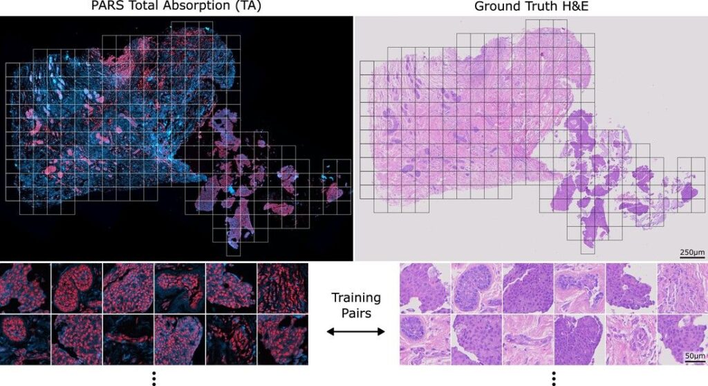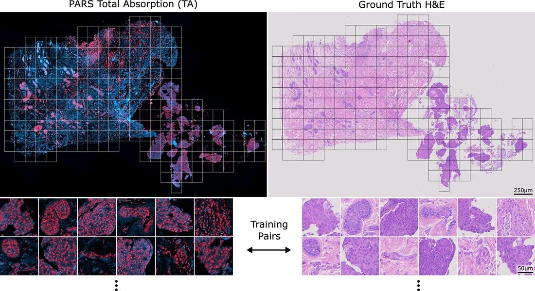
Since the dawn of pathology, we have relied on chemical staining to visualize cellular and tissue details under the microscope. Stains like H&E do not show the tissue as it is, but rather color it in a way that facilitates our distinction between nuclei, cytoplasm, and connective tissue.
However, this approach has limitations: it consumes the sample, is time-consuming, and provides only one type of contrast at a time.
This is where
IllumiSonics MLI – Multi-Laser Imaging comes in:
The Core Idea
The technology relies on multi-color/multi-wavelength laser beams directed at the tissue.
When light interacts with tissue, several natural phenomena occur:
• Some light is absorbed.
• Some is reflected.
• Some changes depending on the type of molecules present.
These interactions serve as a unique optical and molecular fingerprint for each cell, protein, lipid, and tissue.
The device captures these fingerprints digitally with high precision.
How are the results interpreted?
Instead of adding a chemical stain, artificial intelligence (AI) transforms these fingerprints into images highly similar to traditional stains.
In other words:
• We do not physically stain the tissue.
• But we can visualize the tissue as if we had applied H&E, PAS, or Masson’s Trichrome, among others.
Furthermore, MLI can reveal additional information not possible with conventional stains, such as:
• Total Absorption (TA): The amount of light absorbed by the tissue.
• Quantum Efficiency Ratio (QER): The efficiency of the photochemical reaction of molecules within the tissue.
This data reveals different levels of molecular composition, which was not possible in traditional pathology.
A Simple Analogy
Imagine the sample is an uncolored canvas.
In traditional methods, we added colors (stains) to visualize it.
But with MLI, the canvas itself illuminates its natural colors when the appropriate light is shone upon it, and then the computer reconstructs a complete image, which can be switched between H&E, PAS, etc., without altering the original canvas.
What does this practically mean for the pathologist?
• The same slide can yield dozens of virtual “stains.”
• No need for re-biopsy or tissue wastage.
• Faster results: Instead of waiting a long time for staining and examination, you can obtain a diagnostic image within minutes.
• Higher accuracy: Because you see not only the morphology of cells but also their molecular characteristics.
Conclusion:
MLI technology doesn’t just change the staining tool; it transforms how we view the tissue itself:
• From a superficial view with chemical colors,
• To a true molecular view using light.