

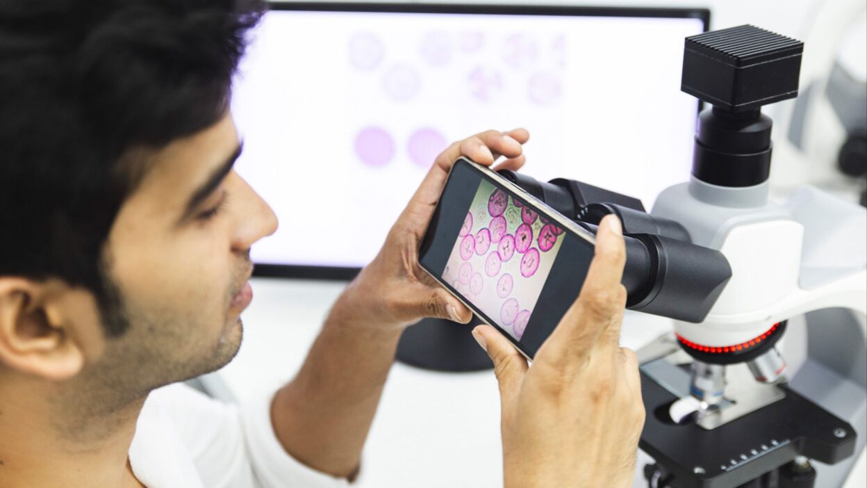
Digital pathology means that a glass slide is no longer limited to the microscope stage. The slide is scanned with a slide scanner and converted into a high-resolution whole slide image, usually called a WSI. A pathologist can then open that slide on a workstation, move through the tissue, zoom in and out, annotate findings, share the case with a colleague, or connect the image to the laboratory information system.
The point is not to replace the pathologist with software. The point is to turn a single fragile glass slide into a visual file that can be reviewed, archived, taught, measured, and shared with far less friction.
How a slide becomes a digital case
The beginning is familiar: tissue processing, paraffin embedding, sectioning, staining, and a glass slide. After that, the slide enters the scanner. The scanner captures many small fields at a defined magnification, often 20x or 40x, and stitches them into one large image of the whole tissue section.
This is not a normal photograph. A whole slide image can be several gigabytes in size. It needs fast viewing software, organised storage, a stable network, and a display good enough for diagnostic review. If one of these pieces is weak, digital pathology becomes frustrating instead of useful.
What the pathologist sees
The digital viewer works a little like an online map. The pathologist can start with the full section, then move gradually to glands, nuclei, margins, necrosis, mitotic figures, or any area that needs closer review. The viewer can also support annotations, measurements, screenshots, and comparison between stains.
These tools do not remove diagnostic skill. They make parts of that skill easier to document and share. Instead of saying that a suspicious area was seen near one edge of the slide, the pathologist can mark the exact region and return to it later.
Where digital pathology helps in daily work
The first clear use is remote consultation. A difficult case can be shared with a subspecialist in another city or country without shipping the glass slide. This matters for hospitals that cannot cover every subspecialty locally, including dermatopathology, renal pathology, neuropathology, and rare tumours.
Archiving is another major use. Glass slides can break, fade, become misplaced, or be difficult to retrieve years later. A digital archive does not solve every storage problem by itself, but it gives the laboratory a searchable copy that can be reviewed again, provided naming, backup, retention, and privacy rules are clear.
Training also changes. Residents and students can study the same slide at the same time, including rare cases that would otherwise be locked in a box. A teaching set can include annotations, key regions, and comparison images. That is a major gain when good teaching slides are limited.
Where AI fits
Artificial intelligence needs digital slides. A model cannot analyse a glass slide sitting in a cabinet. Once the slide becomes an image file, algorithms can help with defined tasks: cell counting, immunohistochemistry quantification, tumour area measurement, region detection, or case prioritisation.
Safe AI use starts with a narrow question. Are we counting Ki-67? Looking for small nodal metastases? Measuring tumour burden? Each task needs local validation against pathologist review. A model that performs well in a publication may behave differently with another scanner, stain, tissue preparation method, or patient population.
Why buying a scanner is not enough
A scanner is only one part of the system. The laboratory still needs a workflow: who loads the slides, who checks scan quality, where the files are stored, how the images connect to the LIS, who can access them, and what happens when storage fills or the network slows down.
Small details matter. Dust, air bubbles, tissue folds, thick sections, staining variation, focus quality, upload speed, and monitor calibration can all affect the experience. A poor glass slide does not become a good case because it was scanned. Digital pathology often makes the original slide quality more visible.
Can digital pathology be used for primary diagnosis?
Yes, but it requires local validation before routine use. The laboratory should compare digital review with microscope review across specimen types, document performance, train users on the viewer and displays, and define which cases still require microscope review.
Some diagnoses are straightforward digitally. Others require caution, including tiny organisms, subtle mitotic figures, crystals, some stains, or slides with uneven focus. The useful question is not whether digital is better than the microscope. The useful question is which cases can be signed out digitally, on which system, and under which safeguards.
Why it matters for resource-limited laboratories
For many laboratories, the immediate value is not AI. It is access. A small laboratory can request a second opinion quickly. A remote hospital can share a difficult case without transporting slides. A training programme can build a local digital library. A referral centre can support several laboratories without physically receiving every slide.
The best implementation is usually gradual: consultation cases first, then teaching sets, then selected diagnostic workflows, and only after that AI tools for specific tasks. This route is safer and cheaper than buying a large platform without an operating plan.
Bottom line
Digital pathology moves the slide into a digital environment where it can be viewed, shared, measured, archived, and taught. Its value comes from workflow, not from the image alone.
Done well, it can shorten consultation time, improve training, support quality control, and prepare the laboratory for responsible AI use. Done without planning, it creates large files, storage pressure, and screens that pathologists avoid. The difference is not the scanner alone. It is how the laboratory builds the work around it.